Problematic Pedestrian Bridges
By Henry Petroski
Crowds walking, running, dancing, and even standing still on these spans often prove to be the ultimate test of a structure's soundness.
Crowds walking, running, dancing, and even standing still on these spans often prove to be the ultimate test of a structure's soundness.

Pedestrian bridges are ubiquitous, but most are unremarkable and anonymous. They span interstate highways to carry hiking and biking trails over heavy traffic, thus keeping walkers, runners, and cyclists safely above the fray. Some pedestrian bridges rise to prominence, however, often because of controversies or problems associated with their location, design, construction, or performance.
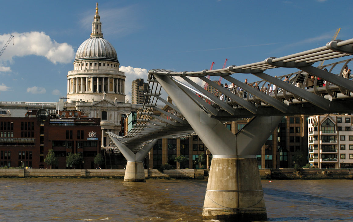
Brian Jeffery Beggerly/Wikimedia Commons
Click "American Scientist" to access home page

American Scientist Comments and Discussion
To discuss our articles or comment on them, please share them and tag American Scientist on social media platforms. Here are links to our profiles on Twitter, Facebook, and LinkedIn.
If we re-share your post, we will moderate comments/discussion following our comments policy.