The Shape of the Universe: Ten Possibilities
By Joey Shapiro
Recent experimental evidence has hinted that the shape of the universe may be found among the ten orientable Euclidean 3-manifolds
Recent experimental evidence has hinted that the shape of the universe may be found among the ten orientable Euclidean 3-manifolds

DOI: 10.1511/2001.34.443
Throughout most of antiquity, people who thought about the shape of the world generally assumed they lived on a vast flat plane—albeit a slightly bumpy one. It was a reasonable belief that weathered thousands of years and almost as many philosophers.
Legend has it that Aristotle, during the fourth century B.C., watched a ship disappear over the horizon—hulls first, then sails, then mast. The ship, he noticed, did not become smaller and smaller, disappearing into nothingness. It sank over the horizon. His conclusion that the earth must be round (supporting with observation and deduction an earlier speculation by Pythagoras) was one of the great intellectual achievements of all time.
Over the intervening millennia, we have discovered many other secrets about our planet, our galaxy and our universe. But a fundamental question remains unanswered. What is the shape of the universe within which we reside?
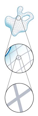
Fortunately, recent observations in astronomy are beginning to hint at the shape of the spatial universe—or at least limit the wide range of possibilities. One type of shape, called a Euclidean 3-manifold, has arisen as a prime candidate. Amazingly, mathematicians have shown that there are only 18 Euclidean 3-manifolds and, of these, only ten are probable candidates for the universe.
We would like to help the reader visualize these candidates for the universe by first describing simpler analogues that can be thought of as two-dimensional universes. Then we will visualize the three-dimensional shapes and discuss how ongoing work in astronomy may help us to finally answer the question: What is the shape of the universe?
Mathematicians who talk about the shape of the universe are referring to its topological shape. In topology, objects are treated as if they are made of rubber. In this medium a doughnut is the same thing as a coffee cup. That is, we can deform a very malleable doughnut into the shape of a coffee cup without any cutting or pasting.

But, topologically speaking, the surface of a doughnut, a torus, is not the same as a sphere, the surface of a solid ball. There is no way to mold one into the other without cutting and pasting.
There are many more surfaces that are topologically distinct from these two. For instance, we can add handles to the torus. Each handle creates a new hole. Thus, the torus, a one-handled surface, has one hole, whereas a two-handled surface has two. Topologically, the number of handles defines the surface. Any two surfaces with different numbers of handles are distinct. With this information we can already generate an infinite number of distinct surfaces.
Tom Dunne
We call all of these surfaces 2-manifolds—they all share a defining property. Around any point on these surfaces exists a disk of points. The disk might be very small and slightly bowed, but its existence tells us that, locally, the surface is two-dimensional.
This definition may sound technical, but we encounter this property every day. From our vantage point on the surface, the Earth looks flat. Locally, the surface of the Earth appears two-dimensional—there is a disk of points around every point on the surface. If we only saw this local picture, it would be reasonable to believe that the Earth is an infinite plane, a sphere, a torus, or any one of the infinite number of multi-handled surfaces.
Some of these topological shapes can be pretty tricky to comprehend, even for topologists. To make visualizing them easier, topologists have developed techniques that simplify the process.
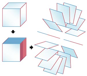
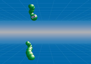
One way to picture a torus is to start with a square, called a fundamental domain for the torus. Pretend the square is a piece of paper, and construct a cylinder by gluing the left side of the square to the right side. The top and bottom sides of the paper have become the circles on the top and bottom of the cylinder. Gluing those two circles together creates a torus.
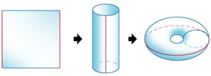
We can keep track of a two-dimensional bug walking around the surface of the torus by watching the bug move around the square. Each time the bug reaches the top edge of the square, transport it to the corresponding point on the bottom edge of the square. Each time the bug walks off the right edge of the square, transport it to the corresponding point on the left edge of the square.
This system for visualizing a torus has two advantages. First of all, we can keep track of an action that is taking place in three-dimensional space, a bug walking on a torus, with a picture in two-space, a bug on a square with some transport features. Secondly, the plane has a nice Euclidean geometry. In Euclidean geometry, the parallel postulate holds; for each line and a point off the line, there is a unique line parallel to the first that passes through the point. And the sum of the angles of a triangle always equals 180 degrees. These statements are not always true in other geometries (spherical and hyperbolic geometries will be explored later). But, because the square is sitting in this Euclidean geometry, we can give its geometry to the torus. We say that the torus is a Euclidean 2-manifold.
Instead of a square, we could form the torus from a parallelogram by gluing its opposite edges together. Although this would not change the topology of the resulting surface, it could change the lengths of the loops that go the short and long way around the torus, and the angles between them. There are an infinite number of possible ways to model a torus as we vary our fundamental domain over the possible parallelograms.
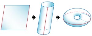
However, we cannot use just any quadrilateral as a fundamental domain. When we glue one edge to another, they must be the same length. We don't want to stretch or contract an edge while gluing—that would ruin the Euclidean geometry we want the surface to inherit.
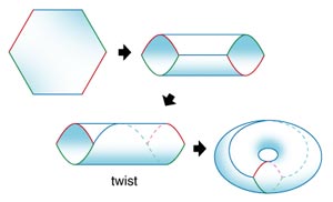
In addition to parallelograms, we can also use a hexagon. By gluing together the opposite edges, we again obtain the torus.
Now we are ready to step up a dimension.
No matter where we have been in the universe, if we picked a point nearby and considered all of the points within a distance of two feet from that point, we then observe a three-dimensional ball of points. Cosmologists believe this is true throughout the universe. That special property leads them to conclude that the universe is a 3-manifold.
But which 3-manifold is it? Trying to determine which 3-manifold describes the universe is a daunting challenge. Mathematicians long ago concluded that, as with 2-manifolds, there are an infinite number of these shapes. In and of itself, that is not a problem. We could still hope to make a complete list of the possibilities, just as mathematicians have done for two-dimensional surfaces. Nobody has yet succeeded in creating such a list. Luckily, there are physical properties of the observed universe that can help limit the possibilities without a complete list. One of these properties, curvature, could have major implications for the topology of the universe.
About 300,000 years after the Big Bang, the temperature of the universe cooled enough to allow electrons and protons to combine, forming the first atoms. When this happened, the radiation now known as cosmic microwave background radiation, which had previously been constantly scattered by free charged particles, was suddenly able to travel, unimpeded, throughout the expanding universe. This radiation is surprisingly uniform, varying only slightly over great distances. Uniformity like this should only occur in a universe whose curvature does not vary with either position or direction.
Tom Dunne
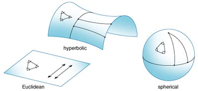
Therefore, the spatial universe is believed to have one of three possible geometries: spherical geometry with positive curvature, Euclidean geometry with zero curvature, or hyperbolic geometry with negative curvature. Two-dimensional analogues of spherical, Euclidean and hyperbolic geometry appear in Figure 2.
These three geometries have very different properties. For example, recall that in Euclidean geometry, the sum of the angles of a triangle adds up to 180 degrees. In spherical geometry this is not the case. If three points are placed on a sphere, the angles between them sums to more than 180 degrees. In hyperbolic geometry, triangles can be formed where the sum of their angles is a positive number strictly less than 180 degrees.
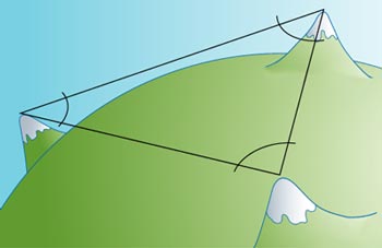
By the first half of the 19th century, Carl Friedrich Gauss understood the possibility that our universe might not be Euclidean. He compared the angles of the triangle formed by three mountain peaks in Germany. Their sum, within the tolerance of error, was 180 degrees. On the small (astronomically speaking) scale that Gauss measured, the universe appeared to be Euclidean.
Tom Dunne
We cannot safely extrapolate Gauss's mountain data to the universe at large. Perhaps the angles of a triangle formed by three distant galaxies do not add up to exactly 180 degrees. Perhaps the geometry of the universe is spherical or hyperbolic but appears Euclidean within the tiny region that we can observe.
A variety of recent experiments, examining everything from supernovae to cosmic microwave radiation, have explored this mystery. A recent study measured the angular power spectrum of the cosmic microwave background radiation using high-altitude balloons over Antarctica. There was a peak in the power spectrum that, the researchers believe, can only be explained by the existence of cold dark matter—relatively large, slow-moving particles that do not emit light—in a Euclidean universe. Other studies have also leant support to the possibility that the universe is Euclidean. Perhaps Gauss was right all along.
If one believes the universe is Euclidean, the number of possible shapes shrinks dramatically. In 1934, Werner Nowacki proved that there are only 18 possible Euclidean 3-manifolds. (W. Hantschze and H. Wendt published a more direct classification in 1935.) Instead of an infinite list of 3-manifolds, we need only consider 18 possibilities for the spatial universe. Understanding the properties and appearances of these manifolds may supply the information needed to determine experimentally the universe's shape.
Of these 18 Euclidean 3-manifolds, eight are nonorientable; they contain an orientation-reversing loop. If you flew from Earth along such a loop, you would eventually return home with your orientation reversed. Your heart would be on the wrong side of your body. Your wristwatch would turn counterclockwise instead of clockwise. At least, this is how you would appear to the other residents of Earth. You would see no difference in yourself. To you, it would appear that you had returned to a mirror copy of the Earth. All of the clocks would run counterclockwise. All of the writing would appear as mirror writing. Every person's heart would appear on the wrong side of his body.
As fascinating as the idea of a nonorientable universe is, it is unlikely that we are living in one. If the universe were nonorientable, cosmologists predict that we would observe energy radiating from the boundary zones where regions dominated by anti-matter and matter meet. This strange interaction has never been observed. Therefore, although it is possible that, if the universe is large enough, these boundary zones exist outside of our field of vision, it is relatively safe to restrict our discussion to the ten orientable Euclidean 3-manifolds.
Three-manifolds are extremely difficult to visualize. We will attempt to simplify this task by always describing the Euclidean 3-manifolds with the technique used to visualize 2-manifolds. Remember that we first used a square as the fundamental domain of a torus. A torus was created when the opposite edges of the square were glued together. Now, when we visualize 3-manifolds, we will use the same technique, but with a 3-dimensional object as the fundamental domain.
Tom Dunne
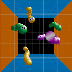
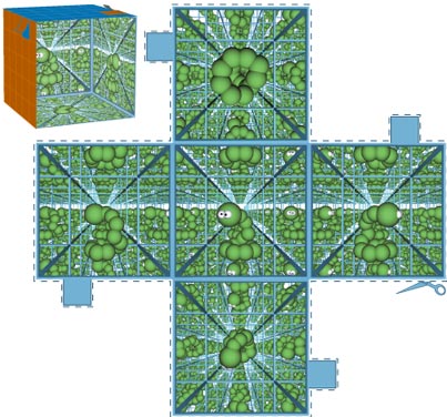
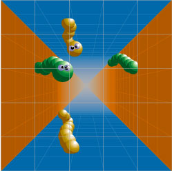
The 3-torus is the generalization of the torus in a higher dimension. Instead of gluing together the opposite edges of a square, the opposite faces of a cube are joined. In the 3-torus, every point on a face of the cube is glued to the corresponding point at exactly the same spot on the opposite face.
If you were somehow in this 3-manifold and looked forward, you would see the back of your own head. You would see copies of yourself in each face of the cube: forward, backward, left, right, above and below. Past these copies, other copies would be visible—copies as far as the eye could see. Standing in a 3-torus and looking out is similar to standing in a fun-house room of mirrors. But in the 3-torus, the images are never reversed (see Figure 5).
Noah Eisenkraft
It is important to note the circular nature of this and many other manifolds. If this 3-manifold were really the shape of the universe, you could fly from Earth in a particular direction and, without ever changing course, eventually return home. This sounds impossible, but a similar phenomenon exists on Earth. If you head due west along the Equator, it is common knowledge that you will one day return to your starting point.
Another interesting property of the 3-torus is its relation to the two-dimensional torus (2-torus) explored earlier. If we cut the cube into tiny vertical slices, we would obtain a series of squares. The opposite edges of these squares would be glued together because those edges composed the opposite faces of the cube. The 3-torus is like a continuous Rolodex, a circle of 2-tori. Remember that the front and back square are connected; they were originally the faces of the cube. Topologists denote this manifold T2xS1, where T2 denotes the 2-torus and S1 denotes the circle. This is an example of a torus bundle, consisting, as it does, of a bundle of tori.
A cube is not the only shape that generates a 3-torus. Just as the parallelogram generated a 2-torus, a parallelepiped (a three-dimensional object with parallelograms for faces) easily generates a 3-torus. By gluing together the opposite faces of different parallelepipeds, spaces with different closed curves and with different angles between those curves are generated.
These, and all other finite manifolds, supply an easy way to picture an expanding universe. If the fundamental domain of a manifold expands over time, then the space it generates will expand with it. Every point in an expanding space is moving farther away from every other point, exactly what we see in our universe. Keep in mind, however, that points near one face will always remain very close to points on the opposite face; opposite faces are glued together, regardless of the fundamental domain's size.
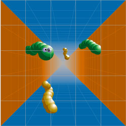
The 1/2-Twist Cube Space is very similar to the 3-torus. The fundamental domain even remains a cube, although parallelepipeds work just as well. Four of the faces are glued the same way. The remaining two, the front and back faces, are glued together, but with a 180-degree twist. The top of the front face is glued to the bottom of the back face. If you were in this manifold and looked out one of these faces, you would still see a copy of yourself, an upside-down copy. Beyond that, a normal, "right side up" copy would be visible, and so forth.
Tom Dunne
Like the 3-torus, the 1/2-Twist Cube Space can be vertically sliced into a deck of 2-tori. This time, however, the front 2-torus is glued to the back 2-torus with a 180-degree twist. The 1/2-Twist Cube Space is also a torus bundle.
The next manifold is the 1/4-Twist Cube Space. This torus bundle is generated exactly the same way as the 1/2-Twist Cube Space, but with only a 90-degree twist, not the 180-degree rotation we just used. Because only a quarter turn is mandated, a random parallelepiped will not always generate this Euclidean manifold. The fundamental domain's front and back faces must be squares to avoid distortion. Staring out the front face of the cube you would see copy after copy of yourself, each one a 90-degree rotation of the preceding copy.
Tom Dunne
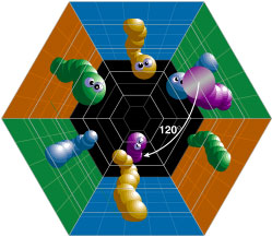
The 1/3-Twist Hexagonal Prism Space, as its name implies, does not use a cube as its fundamental domain. The hexagonal prism that generates this manifold may be less familiar, but is still relatively simple.
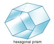
Generating the 1/3-Twist Hexagonal Prism Space is straightforward. Glue each parallelogram directly across to its opposite face. Then glue the two hexagonal faces together with a 120-degree twist. Each hexagonal slice of this manifold is a torus; therefore this too is a torus bundle. If you looked out one of the hexagonal faces you would see that each copy is rotated 120 degrees more than the preceding copy. The copies would not be rotated if you looked into a parallelogram face.
Tom Dunne
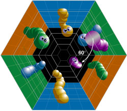
The 1/6-Twist Hexagonal Prism Space is constructed in a similar way to the 1/3-Twist Hexagonal Space. This time, however, the front hexagonal face is glued to the back hexagonal face with a rotation of only 60 degrees. The top edge of one hexagonal face is glued to the second edge of the opposite face. Again, in this torus bundle, the remaining parallelogram faces are glued straight across.
Tom Dunne
The Double Cube Space, or Hantschze-Wendt manifold, is a radically different manifold. This finite space is not a torus bundle and has an unusual gluing pattern. The Double Cube Space, however, still uses a very simple fundamental domain: two cubes, one sitting on top of the other. Figure 10 shows the gluing pattern that generates this manifold. It is important to note that not all of the faces are glued across. Instead, the top front and top back faces are glued to the faces directly below them. In this space, you would see yourself with a peculiar perspective. If you were tall enough, you would see your feet directly in front of your face.
Tom Dunne
With the Double Cube Space the list of finite orientable Euclidean 3-manifolds is complete. It is likely that the shape of the universe lies within these so-called compact manifolds. Many cosmologists believe, for both aesthetic and theoretical reasons, that the universe is not infinite in nature. That assertion makes sense. We all think it's foolish to believe the Earth is an infinite plane—why do we continue to think that the universe is infinite? It would be very difficult to come up with a physical mechanism for the creation of an infinite universe. How would it have started? However, it is still important to consider the four noncompact, orientable Euclidean 3-manifolds until substantial real-world evidence is raised against them.
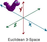
The first, and simplest, of the infinite 3-manifolds is one we are already familiar with. It is  3
3
, Euclidean 3-Space, the space from high school geometry where three axes extend out into infinity. You would see no copies of yourself, twisted or otherwise, if you looked out into Euclidean 3-Space.
The fundamental domain of Slab Space is, unsurprisingly, an infinite slab of space. The top of the slab is an infinite plane glued directly onto the bottom of the slab, another infinite plane. These planes must be parallel to one another but can be rotated or shifted arbitrarily. There is no need to be concerned with these changes because of the infinite nature of the planes—no matter how far we move or rotate one plane, it will always affix perfectly to the other.
Tom Dunne
Topologists use shorthand product notation to describe this manifold. The manifold contains an interval of planes that, after being glued together, form a circle similar to the 3-torus Rolodex. Topologists describe this as  2
2
xS1, where 2
represents the plane and S1, the circle.
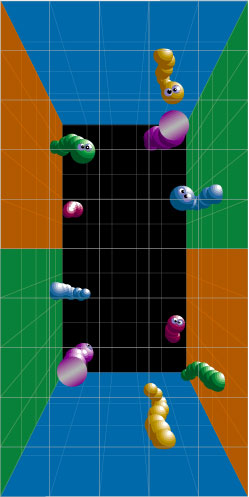
The final two 3-manifolds use infinitely tall chimneys as their fundamental domain. Chimneys are made up of four faces, arranged as the edges of a parallelogram. Chimneys lack both a top and a bottom—their four faces extend infinitely in both of those directions. As with the cubes or hexagonal prism, how this fundamental domain is glued together dictates which manifold is formed.
Tom Dunne
A Chimney Space is formed when both sets of opposite faces are glued together, straight across. After gluing, the parallelogram cross-section is nothing more than a 2-torus. Therefore, topologists refer to this space as the product T2x1.
Tom Dunne
Add a 180-degree rotation to one of the gluings in a chimney space to form a Twisted Chimney Space. This twist, combined with the infinite height of the chimney, provides some unusual characteristics. Examine, for instance, a point, very high up, at one extreme of the Twisted Chimney Space's fundamental domain. Compare that point to another point, a great distance down, at the fundamental domain's other extreme. They seem very far apart. However, after the faces are glued together, those points are surprisingly close.
How can we further whittle down this list of possibilities? Astronomers must gather more evidence and perform more experiments. Mathematicians must develop procedures that take advantage of astronomical data.
The simplest procedure is to look for copies of our Galaxy in the night sky. If we find copies, we can determine the gluing of the universe's fundamental domain. If the universe happens to be a 1/4-Twist Cube Space, un-rotated copies of our galaxy would be visible on four sides, while 90-degree rotations could be seen on the remaining two. Seems easy, right? Unfortunately, this technique holds little promise.
As you know, light travels at a finite speed. Looking out into the universe, we, in effect, look back in time. Even if we someday find an image of our Galaxy, we may not recognize it. Our Galaxy may have looked completely different in its younger years. It would be too difficult, with the sheer number of galaxies out in the universe, to determine which particular one was a copy of our own.
Some cosmologists, after giving up on finding our Galaxy, still hope to find repeating patterns in the sky: copies of quasars, gamma-ray bursts or galaxy clusters. Others have pioneered new ways to attack the problem.
Earlier in this article it was concluded that the universe had a constant curvature. The uniformity of cosmic microwave background (CMB) radiation strongly suggested this. Remember, however, that the CMB has slight variations, tiny changes on the order of 10–5 kelvins.
These small variations in the CMB show us the minute density differences of the early universe. When the universe cooled and expanded, the extra density in these areas slowed these areas' expansion. This effect compounded, eventually clumping the region's matter into galaxies, stars and planets. Looking at a map of the CMB allows us to look back in time, past this intergalactic clutter, at the original density differences. We look at the blueprints of the universe, blueprints that are less than one-thousandth the size of our present-day universe.
Tom Dunne
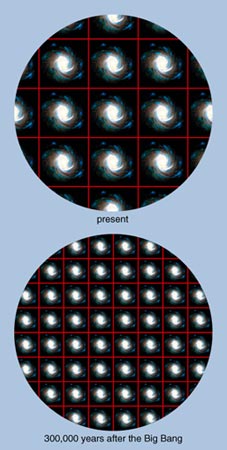
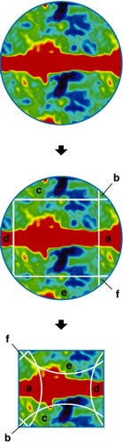
To more readily understand the potential of CMB maps, it is best to consider the example of a two-dimensional torus universe. In the upper panel of Figure 14, you can see how that torus universe "looks." The large squares show the fundamental domain of the torus repeating, with a two-dimensional "reference" galaxy appearing within each copy. The bottom of the figure shows the two-dimensional universe 300,000 years after the Big Bang—what a CMB map will show us. The torus universe would appear smaller, so the copies of the square are smaller in the CMB. Mapping the CMB of the two-dimensional universe, or our own, creates a snapshot of the past.
In a three-dimensional universe we observe all of the spherical sky. Residents of a two-dimensional universe would all be two-dimensional creatures, only able to observe a circle's worth of CMB information at one time. If the circle of temperature variations that they were able to see was smaller than the universe's fundamental domain, they would have no indication of the shape of the universe. If, however, their circle of vision were larger than a single fundamental domain, the creatures would see an intersection. More importantly, they would see patterns repeat. Remember, every square shown is identical, a copy of the fundamental domain.
Once the two-dimensional creatures looked into their visual circle, they could try to find points with matching temperatures. If there were two different points on their visual circle with exactly the same temperature, the points might correspond to the same area in the universe. If there were enough matching temperatures in the two-dimensional creatures' visual circle, they could conclude that they were living in a torus universe.
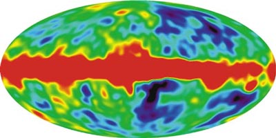
Image courtesy of the Microwave Anisotropy Probe Science Team, NASA
We, however, live in a three-dimensional universe. We observe a sphere's worth of information. And yet, when we map out the CMB variations, we confront the same problem as the two-dimensional creatures. If our sphere of vision is smaller than the fundamental domain of the 300,000-year-old universe, we discover nothing. If, however, our sphere of vision is larger than the CMB universe's fundamental domain, then the sphere will overlap itself, intersecting along circles.
If those overlaps occur, cosmologists will search for patterns in the temperature variations. If there are two circles on the sphere that have the exact same sequence of CMB variation, the cosmologists can compare the circles' orientations. If the circles match up directly across, there is a gluing but no twisting. Some, however, may match after a quarter-twist, or a half-twist. If enough of these matching circles are found, the fundamental domain of the universe and its gluing pattern will be unearthed.
Cosmologists, however, will have nothing to study until a precise temperature map of the CMB is generated.
Noah Eisenkraft
In 1989 NASA sent up its first try: the Cosmic Background Explorer, a satellite that has since completed a temperature map of the CMB from space. Unfortunately, the satellite's angular resolution, on the order of 10 degrees, was not fine enough to make the precise measurements cosmologists needed.
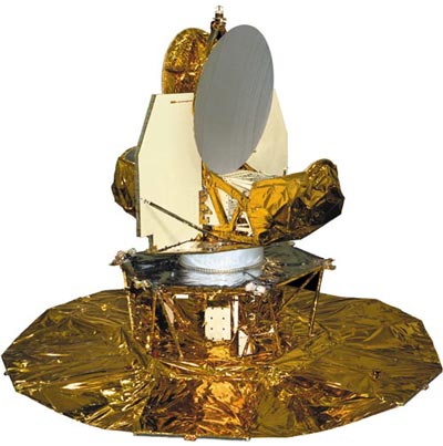
Image courtesy of the Microwave Anistropy Probe Science Team, NASA
This spring, NASA began its second attempt and launched the Microwave Anisotropy Probe. This satellite will map out the temperature fluctuations in the CMB with angular resolution on the order of 0.2 degrees, a vast improvement. Finally, in 2007, the European Space Agency plans to map these fluctuations with the Planck Satellite. It has an angular resolution of 5 arcseconds, 144 times more powerful than the Microwave Anisotropy Probe.
If these satellites are successful we will have precise CMB maps in four to ten years. If our sphere of vision is large enough, if our measurements are accurate enough, and if our data set is good enough, we will know the shape of the universe. Aristotle would be pleased.
Click "American Scientist" to access home page
American Scientist Comments and Discussion
To discuss our articles or comment on them, please share them and tag American Scientist on social media platforms. Here are links to our profiles on Twitter, Facebook, and LinkedIn.
If we re-share your post, we will moderate comments/discussion following our comments policy.