No Evidence For New Adult Neurons
By Shawn Sorrells, Arturo Alvarez-Buylla, Mercedes Paredes
Adult human brains don’t grow new neurons in the hippocampus, contrary to the prevailing view.
Adult human brains don’t grow new neurons in the hippocampus, contrary to the prevailing view.
When our recent study of human hippocampal neurogenesis was met with significant skepticism, we weren’t surprised. After all, we ourselves remained skeptical of what we were seeing throughout our investigation. But repeated and varied experiments convinced us that our conclusions were correct: New brain cells don’t grow (or are extremely rare) in the adult human hippocampus, a region important for learning and memory. The birth of new neurons in human memory circuits, in other words, declines during childhood to undetectable levels by adulthood.
Our research findings sparked healthy debate because for about 20 years, brain scientists have thought that neurons continue to be born in the adult human hippocampus. The question of whether and how new neurons are born in adults is important for understanding how our brains adapt to changing life circumstances and how we might be able to repair brain injury.
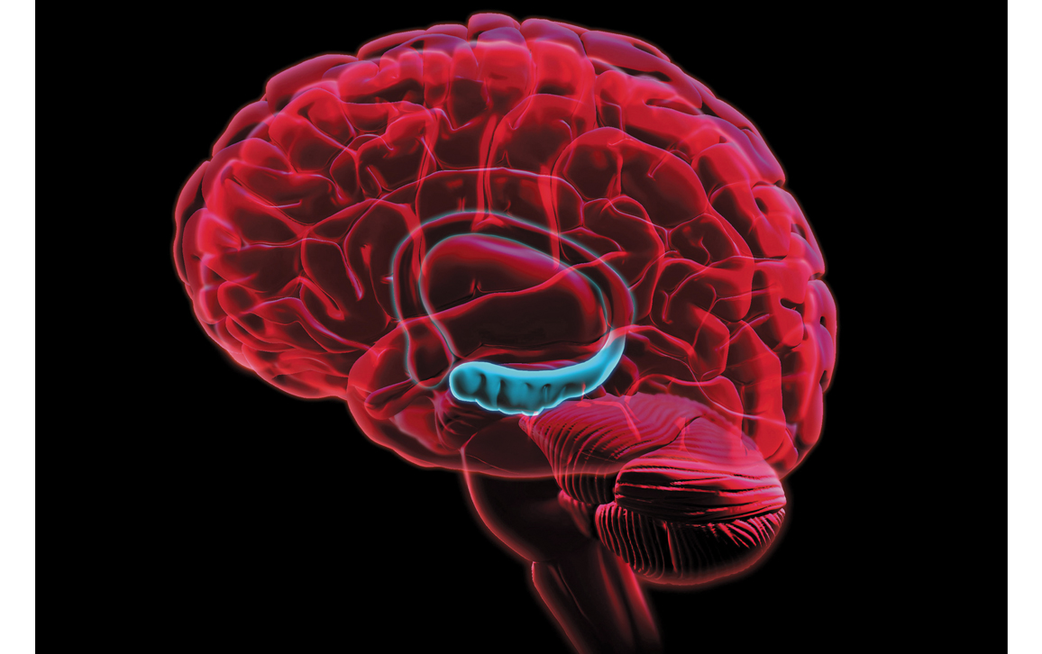
Evan Oto/Science Source
Science advances with the collection of more evidence that helps refine and revise theories. As neuroscientists, we too are adjusting our ideas of how adult human learning must work in light of our recent study.
One of us, Alvarez-Buylla, has been studying how new neurons are born and integrated into brain circuits since the 1980s. He was a member of Fernando Nottebohm’s laboratory at the Rockefeller University, which was at the time producing a groundbreaking series of papers showing that the brains of songbirds grow new neurons each season as they get ready to learn new songs. Earlier research from the 1960s had found evidence that rodent brains produce new neurons during adulthood, but this idea remained highly controversial until Nottebohm’s songbird studies convinced most neuroscientists that adult brains could make new neurons.
NICHD/I. Williams, CC BY 4.0
Since then, several studies have found signs of new neurons in the adult human hippocampus, leading many researchers to accept that this part of the brain could renew itself throughout life in people too. The idea stimulated interest in figuring out how to increase this regenerative capacity and perhaps stave off age-related declines in brain function.
Indeed, we began our search for newborn neurons in the adult human hippocampus because previous studies had estimated that 700 new cells are born in the adult hippocampus per day. We wanted to contrast this finding with results in another region of the brain, where we had recently reported finding far fewer new neurons than seen in other animals.
The first sign that something different might be occurring came when one of us, Alvarez-Buylla, visited the lab of our collaborator Zhengang Yang at Fudan University in China to study several well-preserved human brain specimens. They were not able to detect any new neurons in the adult hippocampus at all.
When Alvarez-Buylla returned from China to our laboratory and shared the observation that new neurons were missing from the adult human hippocampus, we were faced with a challenge: How do you prove a negative? How could we be sure that we weren’t just missing the new neurons that other studies had seen?
How do you prove a negative? How could we be sure that we weren’t just missing the new neurons that other studies had seen?
As some critics have pointed out, identifying new neurons in human brain tissue is complicated. Typically, researchers look for the presence of certain proteins that we know are produced by young neurons. But we were looking at donated brain samples from dead people; maybe these “identifier” proteins degrade after death. They may also have other roles and be produced by other kinds of cells.
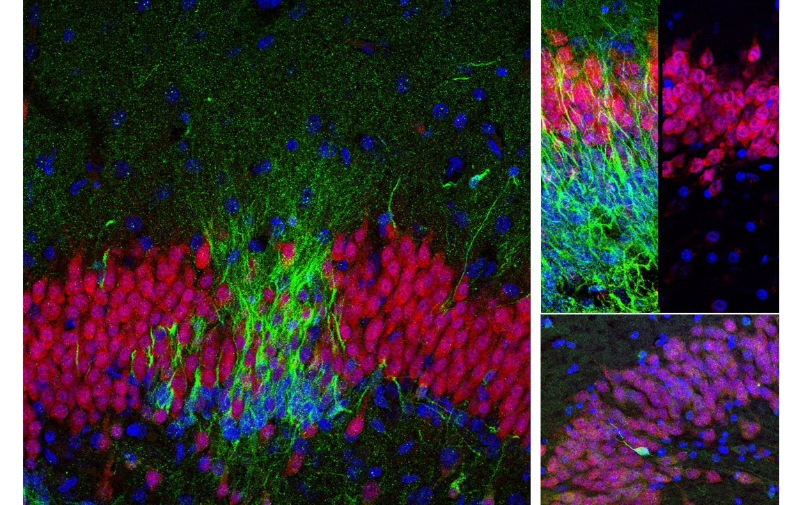
So we needed to use multiple approaches to look for new neurons. First we examined several different proteins that are present in young neurons. We next studied the cells closely with high-resolution light and electron microscopes. We wanted to be sure that any cell we reported would have the distinctive appearance of young neurons; they tend to have a simpler shape that differentiates them from mature neurons, which are usually bigger, with long, elaborate branches. We also looked at overall patterns of gene expression in this region and observed a similar decline in genes associated with young neurons. In addition, we looked for evidence of the stem cells that make young neurons, which have their own protein markers and can be detected when they divide.
None of the adult hippocampal tissue we examined with these techniques showed evidence of young neurons or their dividing stem cell parents.
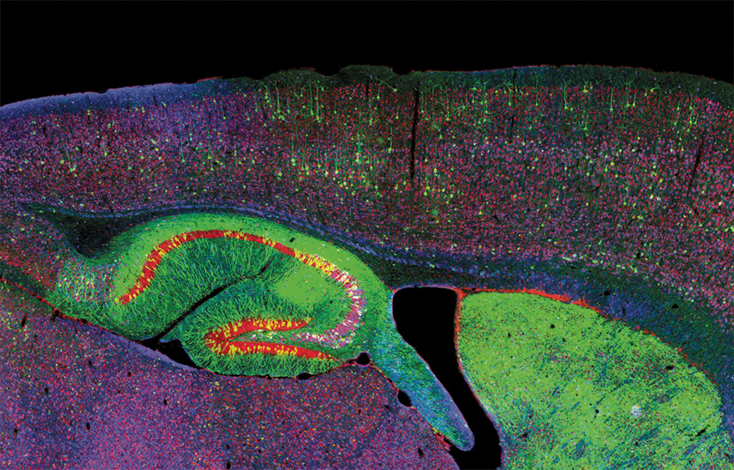
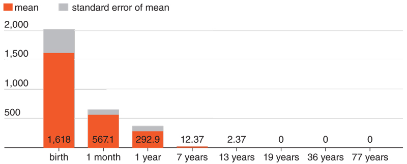
To make sure that our techniques were even capable of detecting young neurons or dividing neural stem cells, we looked at the same region of the hippocampus before birth, when we knew they should be present. In these fetal brain samples, we clearly saw plentiful new neurons. Using the same techniques, we then looked for these cells in brain tissue from people who died in infancy, childhood, or early adolescence. We saw that the number of new neurons sharply declined until few were found in 13-year-olds; by 18 and 19 years of age, we could not find any. If neurogenesis continues in the adult human hippocampus, it is a very rare phenomenon.
Could our inability to see these cells be because of unknown differences between young and old brain tissue? We knew that there are very rare young neurons in other parts of the adult human brain, so we looked in those regions. When we readily found those rare young neurons, we became more confident that what we were seeing, or not seeing, in the hippocampus was not simply an artifact of aging brain tissue.
Could something about the patients’ history prior to death, or the way the samples had been collected, have obscured evidence of new neurons that had been present when the brains had been alive? To convince ourselves that the tissue was as representative of adult brains as possible, we studied brains collected by many different collaborators around the world and saw the same results.
Sorrells et al.
Could the time between death and preservation of the brains lead to our inability to detect young neurons? To test this question, we collected more than a dozen tissue samples from patients who were having brain tissue removed as part of surgical treatment for severe epilepsy. These are samples we collected and preserved quickly to maximize their quality. In addition, we looked at two samples where the brains had been collected and preserved almost immediately at the time of death, and we saw the same results.
In total we examined 59 brains, a collection comparable in size to those examined by previous studies. In all these cases, we saw the same results: no signs of new neurons in the adult hippocampus. We concluded that if new neurons were being born in the adult human hippocampus, then they are extremely rare.
The Conversation/Sorrells et al, CC BY-ND
So what have other researchers seen that made them believe that new neurons are born in the adult human hippocampus? Previous studies frequently used only a single protein to identify new neurons. Unfortunately, we found that the protein most often used, one called doublecortin, can also be seen in nonneuronal brain cells (called glia) that are known to regenerate throughout life.
One other research group tried a different technique more commonly used by archaeologists and geologists: carbon-14 dating. This is a very creative way to determine the age of cells, especially in a field where we need new ways to study the human brain. However, it’s not clear how precisely this method can identify neurons. Nor is it clear whether there are other reasons the radioactive carbon levels might change beyond the cell division that would lead to new neurons.
Our research left us with a lingering question: Why does this decline in neurogenesis happen? Why does the hippocampus continue to create new neurons into adulthood in other animals, but not in humans?
To wrap our heads around this question, we examined the hippocampi of macaque monkeys, which are known to continue producing new neurons into adulthood. Using labeling techniques that are not typically used in humans for ethical reasons, we tracked the generation of new neurons in living animals. We discovered that the neural stem cells that generate new neurons coalesce into a ribbon-like layer in the monkey hippocampus before birth. This layer was present and contained dividing cells even in juvenile monkeys. When we looked back at our data from the newborn human hippocampus, we saw that the stem cells did not organize themselves in this fashion—a clear developmental difference between human brains and those of other primates.
Mark McClendon, Zaida Alvarez Pinto, Samuel I. Stupp, Northwestern University, Evanston, IL, CC BY-NC
Our study only pertains to the hippocampus; many other regions in the human brain—which is very big—have not been investigated and remain to be explored for the possible presence of new neurons. The development of better methods of directly studying the human brain will help researchers understand more about how plasticity occurs in the human hippocampus. And future research can work to determine whether there are ways to reignite the birth of new neurons in this region.
But what does our finding mean? Should we lament the lack of new neurons in the adult human hippocampus? We think not.
First, the process of making a new neuron is fascinating and is already teaching us many new things. Adult neurogenesis should continue to be an area of study in birds, mice, rats, and other species where it occurs. One day this work might teach us how to induce it in the human brain.
Second, our brains operate for decades—much longer than the mouse brain, despite the rodent’s plentiful new neurons. Indeed, the long lives of humans may be linked to the decline in hippocampal neurogenesis; we might run out of progenitors in childhood.
Our work also raises new questions, because clearly a rich and healthy lifestyle does improve human brain function and holds back the decline of age, even without new neurons. Developing a deeper understanding of human brain development may yet provide new treatments and therapies for brain diseases of aging.
This article has been adapted from one that was published on The Conversation, http://theconversation.com. Licensed through the Creative Commons, https://creativecommons.org/licenses/
Click "American Scientist" to access home page

American Scientist Comments and Discussion
To discuss our articles or comment on them, please share them and tag American Scientist on social media platforms. Here are links to our profiles on Twitter, Facebook, and LinkedIn.
If we re-share your post, we will moderate comments/discussion following our comments policy.