Visualizing Biological Networks as Mandalas
By Caryn Babaian
With an Eastern spiritual symbol and some colored chalk, a biology teacher explores the interdependence and the evanescence of all living things.
With an Eastern spiritual symbol and some colored chalk, a biology teacher explores the interdependence and the evanescence of all living things.
DOI: 10.1511/2015.116.322
As anyone immersed in the life sciences can tell you, the world of biology contains a great many interacting systems, but very few of the connections among them can be portrayed simply as straight lines. The more closely we look, the more apparent it becomes that living systems on any scale are complex, self-replicating, and dependent on one another—whether for sustenance, to regulate their growth, or simply to make use of one another’s waste products for their own purposes. From molecular cascades to entire biomes, the connections within and among biological networks multiply and give rise to emergent properties, which in turn foster new connections that develop into networks. Yet most standard introductions to biology omit this dynamic perspective. The conventional classroom practice of presenting material in a linear sequence fails to reflect the ways in which a living planet both produces and consumes energy in a continuous cycle.
Photographs by Caryn Babaian.
With our increased understanding of the complex networks that add up to an evolutionary and ecological framework, we need to teach biology in a way that takes account of this interdependence. The formula for presenting such material, however, is not obvious. For some time, as a teacher of biology, I had been asking myself how to depict the limitless diversity and interactivity of living cells. Was it possible to visualize a process such as symbiosis, or to shape the evolution of bioluminescence into a narrative? What would the coevolution of angiosperms and insects look like if we could show how its spatial and temporal elements have acted upon one another? We already have phylogenetic trees, and we can create circular genomes, but where are we to find the visual tools that will help students see the multilayered, networked events of life? In the end it was not my training in biology but my background in art that led me to just the right visual form: the mandala.
The word mandala in Sanskrit means “circle,” or, as many interpret it, “sacred circle.” As a symbol it represents the whole universe, and even viewed simply as an image the mandala tells a story at many levels. Concentric rings of different colors, for example, may each stand for an element of life such as air or water, with various life forms represented by smaller shapes within the rings, each one distinct and yet related to the others. A few years ago, while teaching physiology in an acupuncture institute, I became fascinated with the Buddhist concept of the mandala and went on to observe the creation of sand mandalas by Tibetan monks. Borrowing the spirit of their creative work, and acting on my innate tendency to use art as a tool for understanding nature, I developed what I came to think of as a biological mandala.
Photographs by Caryn Babaian.
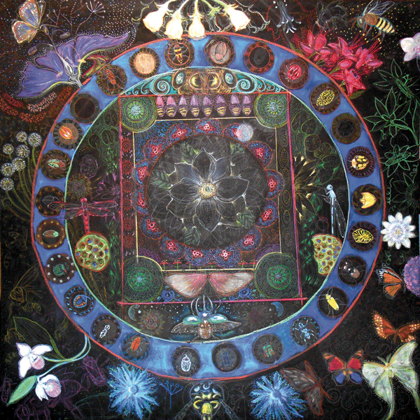
For my first challenge, I tackled the formidably complex concept of the coevolution of insects and angiosperms, or flowering plants. This is a nearly 100-million-year-old story of interdependency and the emergence of whole orders of arthropod pollinators and seed plants. How would I represent intricate relationships, mergers, morphological changes, structural anatomy, extinctions, and adaptive radiations?
As I pored over the fossil evidence and the macroevolutionary patterns (the morphology of plant anatomy, the minutiae of insect feeding parts), the circular format of the mandala started to assist me in organizing the information. In the center I started with the magnolia, one of the oldest flowering plants. Then, progressing through evolutionary time as I worked outward from the center, I began to switch images from early insects (incomplete metamorphosis) to early angiosperms and to include some interactions, including a circle of beetles, the coleopterans, with empty spaces to indicate extinction and new niches.
In a remarkable facsimile of adaptive radiation itself, my biology mandala gained in complexity as new forms of life appeared, diversified, and formed their own connections in the circular network. What had formerly been a textbook lecture now appeared as an ongoing drama, with themes like background extinction, expansion into new niches, and—toward the outer edges, in the present day— human influence and current ecological issues each exerting an impact on the whole system. By envisioning the material as a mandala, I was able to show ecological relationships, evolutionary change, and the visual beauty of the species themselves all together, in a way that scientific illustration rarely even attempts. Moreover, the process of thinking through this visual metaphor further deepened my respect for living systems, which is what good biology teaching should do.
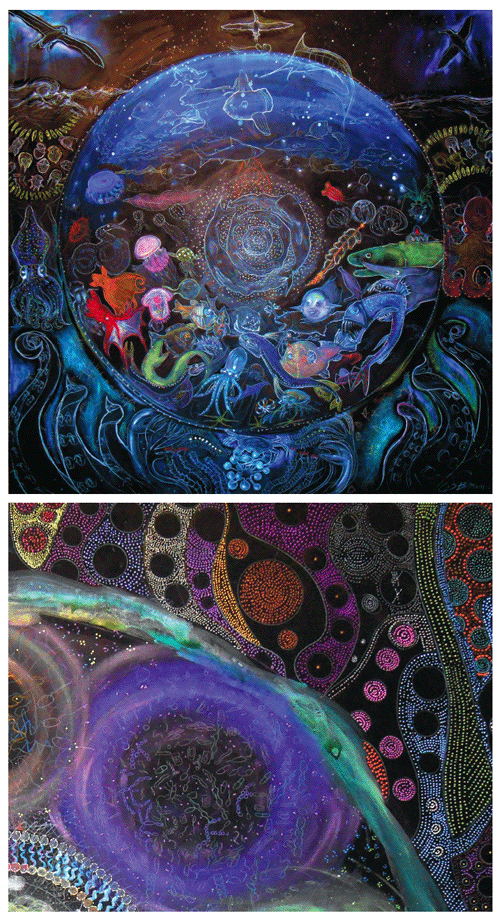
In the center of another mandala, titled “Predators,” I placed the classic predator–prey relationship of the Canadian lynx and the snowshoe hare. We see the lynx, as usual, chasing the snowshoe hare; but in that simple illustration, the hare is also chasing the lynx. The concept of mutual dependence, illustrated by the circle, invites discussion of how predators fill a valuable role in maintaining and regulating ecosystems. This makes more sense to students than the lines on a graph that are typically used to illustrate the predator–prey relationship over time. In the mandala version of this story, the lynx is the ultimate conservationist, with a record of fine-tuning the prey species over millions of years. Healthy predators make for adaptive pressure on their prey, the pressure spreads throughout the ecosystem, and the effects rebound back to the health of the predators.
Yet another mandala (not shown) contains many diverse life forms of the wetlands, from waterfowl to pussy willows, amphibians, and freshwater crustaceans. Although wetlands today remain important both as temporary basins contributing to groundwater recharge and as a reminder of our own phylogenetic transition from aquatic to terrestrial life, they are rapidly disappearing in the face of urban development.
View eight of Babaian's mandalas, and listen to her explain the process of viewing them:
So far I have created eight biology mandalas, and I expect to make more because there are so many biology stories to be told, both inside the classroom and beyond it. For each mandala, my process requires several steps. I start by researching the organisms I plan to depict: reading peer-reviewed papers, revisiting nature journals I had written and illustrated years ago on similar topics, examining zoology specimens and books from over a century ago. Next I walk through the woods, make notes, and move between the science we are taught and the patterns we intuitively relate to. Only after this long incubation process—once all the focal points of biological energy have begun to connect like constellations in the night sky, revealing an underlying unity—can the drawing begin.
The French painter Paul Cézanne may have had a similar process of composition in mind when he said, “Art has a harmony which parallels that of nature.” Cézanne’s observation may be simply another way of stating that art is an emergent property of a creative, living system, embedded within another living system of enormous and unfathomable creative potential.
For me, a biology teacher and an artist, the process of making art has always been a necessity. In the classroom I have used art to help students encounter biology both through their own direct experience (for example, by drawing living forms) and by observation (in narratives about other life forms). Both life-science research and the teaching of biology can benefit immensely from the visual-spatial tool of drawing, which allows us to explore worlds we cannot see or touch.
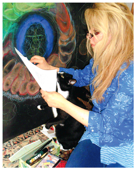
Photograph by Story Biddle.
Using chalk as a medium has allowed me to move easily from one form to another, animate a topic, and then erase it. The erasing is another lesson for me and for students, carrying the message that living systems exist in transient states: Cells divide and die, extinction happens, entropy takes over, new energy flows in, and the system is transformed. In the chalk portrayal of these stories, even the chalk itself has an origin story.
I try to practice evolutionary and ecological-based education, asking students to see themselves as part of our planet’s ecosystem. In learning to draw biological forms the students grow more fully aware of them and become invested in their stories. If they consider their own physiology to be linked with the physiology of the Earth, they can go on to experiment with new ways of creative scientific thinking, and they often become much more invested in caring about the planet as well. It is interdependency that helps to build a healthy ecosystem; if this network of relationships is disrupted, it cannot be easily fixed or replaced. That’s an important lesson for us that I can teach through a biology mandala.
Within the structure of a classroom or the comfortable confines of a living room, it’s almost impossible for most people to fathom the diversity of life forms and the intricacy of the networks connecting them—or to see how losing even one life form would affect the whole. The huge expanses of land and water on the Earth, the extent of evolutionary time scales, and the dynamic qualities of life merging and reemerging, transitioning and transforming, can simply be overwhelming. The mandala, a synthesis of many distinctive elements, helps organize that information. Extinction rates can be expressed in numbers but are not fully appreciated, just as biodiversity and intact ecosystems cannot be fully appreciated unless we see them whole. The apparent chaos and complexity of life are presented in this framework, simplified into a pattern, and structured to represent biological webs, which are neither chaotic nor linear.
Although my audience may enjoy my finished pictures most, I encourage everyone reading this article to take part actively in studying living systems by drawing as you learn. In the process of synthesizing different kinds of information, concepts, and formulas into a cohesive image, you will learn even more. Our modern lifestyle and day-to-day experiences have a tendency to convince us the world is run by cars, products, and buildings and not by biogeochemical cycles, species diversity, photosynthesis, and microorganisms. It’s worth remembering that drawing, like any human adaptation, is a product of those complex living networks and is itself an evolutionarily complex system for exploring our world.
Click "American Scientist" to access home page
American Scientist Comments and Discussion
To discuss our articles or comment on them, please share them and tag American Scientist on social media platforms. Here are links to our profiles on Twitter, Facebook, and LinkedIn.
If we re-share your post, we will moderate comments/discussion following our comments policy.