Salt Marshes Under Siege
By Mark Bertness, Brian Silliman
Agricultural practices, land development and overharvesting of the seas explain complex ecological cascades that threaten our shorelines
Agricultural practices, land development and overharvesting of the seas explain complex ecological cascades that threaten our shorelines

DOI: 10.1511/2004.45.54
An attentive observer sees the richness of life during a single walk along a salt marsh. Gulls circle in the air, and mute swans swoop in to land along the marsh's edge. Dense stands of grasses and other plants—waving gently in the ocean breeze—cover seemingly every spot of ground, from the shore's edge to the tree line. When the tide flows out, crabs scurry back and forth along the muddy edge. In spite of this rich diversity and lush plant life, danger lies ahead for all salt marshes, especially those along the eastern coast of North America.
A salt marsh develops when salt-tolerant plants colonize shoreline sediments on wave-protected shores. The plants bind the sediments, and the vegetation retards water movement and promotes further sedimentation. In New England, a salt marsh creates a series of four strips of habitat. Along the shore, which floods twice most days, cordgrass (Spartina alterniflora) dominates plant life. As ground level rises, marsh hay (Spartina patens) takes over in the so-called seaward edge of the high marsh. At the terrestrial edge of the high marsh, black rush (Juncus gerardi) dominates the landscape. Finally, a band of shrubs marks the inland-most stripe of salt marsh, usually against a line of trees.
Beyond beauty, salt marshes serve as integral components of coastal ecosystems. A salt marsh filters water—from the surface and beneath the ground—as it runs off from terrestrial environments, and that process blocks some nutrients and pollutants from entering estuaries. The dense plants in a salt marsh also protect a coastline from erosion and storm damage. Moreover, many commercially and recreationally important species—including shrimp, oysters and crabs—start life along the edge of a salt marsh, which serves as a marine nursery.
Today, these fertile ecosystems struggle to survive. As this article reveals, a trio of troubles—high-yield agriculture, development and over fishing—threatens salt marshes along much of the eastern coast of the United States and elsewhere. Even now, repairing much of the damage would require the help of nature over thousands of years.
Despite the ecological importance of salt marshes, humans abuse them. Two millennia of livestock grazing and agriculture have made European marshes managed environments rather than natural systems. In New England, early colonists turned livestock loose to graze on marshes and cut marsh hay. In fact, the colonists drained, ditched and diked marshes to enhance the production of this hay, which almost certainly resulted in the loss of considerable plant diversity.
Raymond Gehman / Corbis
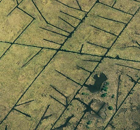
Evidence for this loss of diversity persists. In northern New England, colonists did little ditching and draining of salt marshes, which consist of large areas of intermediate elevation with low plant cover and high plant diversity. In southern New England, on the other hand, colonists ditched and drained almost all marshes, which now consist primarily of dense monocultures of competitively dominant turf grasses. The ditching, however, extended beyond colonial times. In the 19th and 20th centuries, insect control set off more ditching, which further reduced plant diversity. Moreover, developers filled or "reclaimed" many northeastern marshes along the shoreline. All of this human disturbance destroyed an estimated 50 to 70 percent of the marshes that were present in southern New England just before Europeans colonized North America.
Even more trouble lies ahead for salt marshes. Global warming will increase surface temperature, which is predicted to increase soil evaporation. The evaporation could elevate soil salinities in marshes, which will decrease marsh-plant productivity and increase the size, extent and latitudinal range where soil salinities are too stressful for most plants to live.
Global warming also causes thermal expansion of water, which will raise sea level, with some help from the melting of land-based ice sheets. Over the past 50 years, increases in sea level have already gradually shifted the distribution of marsh plants to higher elevations. As sea level continues to rise, and particularly if the pace increases, salt marshes will drown if they are unable to move inland quickly enough. If marshes drown, the loss of large areas of coastal marshes could have catastrophic consequences on coastal systems—increased rates of shoreline erosion, increased nutrients and pollutants from runoff entering estuaries, decreased nursery habitats—as is already occurring in northwest Canada on the shores of the Beaufort Sea. In many cases, the salt marshes will be trapped between rising water on the seaward side and private property and recreational development on the other.
Although global warming and a rising sea level threaten the future of salt marshes, present dangers took hold well in the past. In the 1800s, European merchant ships packed materials and ballast tanks with a reed of the genus Phragmites. In unpacking goods and reducing ballast, ships spread the reed all the way to New England. This reed, though, was no new plant to New England salt marshes. It could be found there 10,000 years ago, but only as a minor member of the plant community along the terrestrial edge. High salt levels push this reed away from the sea. So it grew mostly in freshwater and brackish marshes. Where full-strength seawater invaded a marsh with the rising tide, Phragmites stayed away. Furthermore, dense stands of marsh hay and black rush in typical New England high marshes proved too competitive for the reed to push any closer to shore.
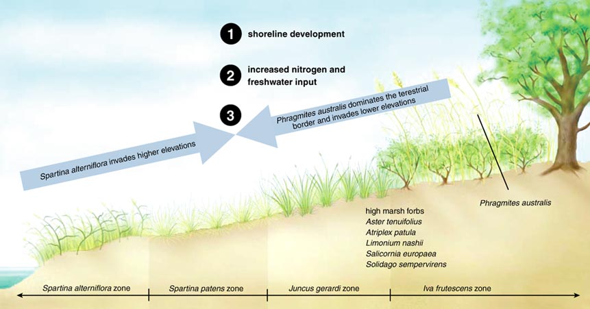
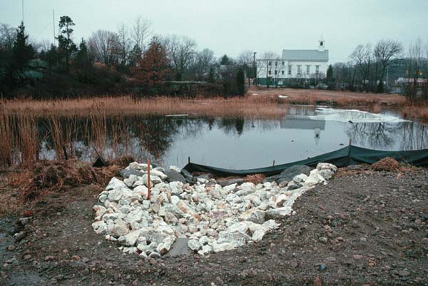
Then, developers changed the character of New England salt marshes by removing the woody vegetation that borders a marsh's terrestrial edge. That vegetation normally blocks runoff water from reaching a marsh. With that vegetation gone, nitrogen-rich freshwater enters a marsh. The salinity of the soil drops and its nitrogen content increases. By lowering salinity, shoreline development removes the major physical barrier that stops Phragmites from migrating close to the saltwater. Similarly, the increased nitrogen supply to the marshes contributes to the shift in the competitive dominance in the high marsh from the native clonal turfs—marsh hay and black rush—to Phragmites.
The reason for this shift is simple. Historically, New England marshes were nitrogen-limited. Marsh hay and black rush, however, capture high levels of the available nitrogen through their roots. Consequently, they dominated the high marsh landscape. When nitrogen is not limiting, the competitive balance shifts to plants that are superior competitors for light, the limiting resource when nutrients are abundant. As the tallest plant in these habitats, Phragmites wins that battle.
In addition, the development-related increase in nitrogen favors cordgrass on the shore side. In historically low-nutrient marshes, marsh hay and black rush outcompete cordgrass, pushing it to the physically stressful lower marsh habitats. With nitrogen loading, however, cordgrass moves to higher elevations, displacing high-marsh perennials.
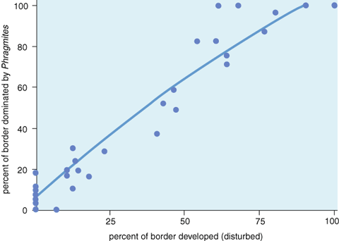
A striking relationship exists between shoreline development and the salt-marsh dominance of Phragmites in southern New England. Two of the authors (Bertness and Silliman) and Patrick Ewanchuk of the Marine Science Center of Northeastern University showed that shoreline development explains over 90 percent of the dominance of Phragmites in these systems. As a result of development, Phragmites started taking over salt marshes from Virginia to Maine during the past 50 years. Although development explains most of this reed's dominance, the 19th-century merchant ships played a role as well, as shown by Kristin Saltonstall of the University of Maryland Center for Environmental Science. She surveyed Phragmites populations across North America and Europe and found that the strain of Phragmites responsible for the expansion in North American marshes is an invasive European genotype—the one used for ballast and packing.
Overall, local shoreline development in New England turned salt marshes from diverse environments to ones dominated by just two plants. This leads to an 80 percent decrease in marsh-plant diversity. The decreasing diversity of plants also reduces the value of a marsh as a nursery for fish and shellfish. These changes can be seen today in salt marshes from Maine to the Chesapeake Bay.
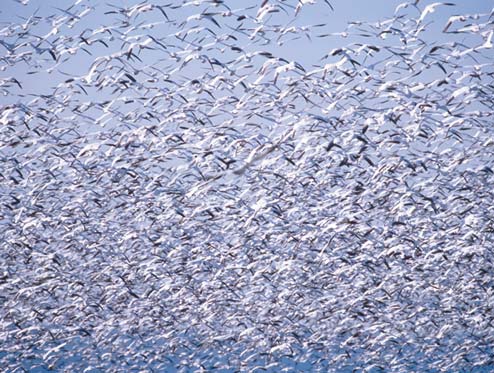
The Hudson Bay Lowland—one of the world's largest wetlands—covers about 350,000 square kilometers, about twice the area of all of New England. This wetland lies south and west of Hudson and James bays. At coastal sites, the Hudson Bay Lowland commonly includes salt marshes, which are highly productive ecosystems by Arctic standards. Each spring, many species of shorebirds, ducks and geese, including the mid-continent population of lesser snow geese, migrate from their wintering grounds in the United States and beyond to breed in these marshes. The lesser snow geese, for example, breed in colonies in the coastal lowlands of Hudson Bay, on Baffin and Southampton islands, and in Queen Maud Gulf in the central Canadian Arctic.
© Owen Kanzler
Lesser snow geese can exert a strong positive impact on the net primary productivity of salt-marsh communities. Like most other salt marshes, ones in the Hudson Bay Lowland are nitrogen-limited. Adult lesser snow geese and their goslings graze intensively on intertidal grasses and sedges during the summer, before migrating along the Mississippi and central flyways in the autumn to spend the winter in Texas and Louisiana. The snow geese live in colonies and adult birds defecate, on average, once every four to five minutes when feeding—enough to cover 40 square meters in a week. The soluble nitrogen in the feces quickly leaches into the soil, where the marsh grasses and sedges capture it. As a result, the geese-grazed plants rapidly regrow.
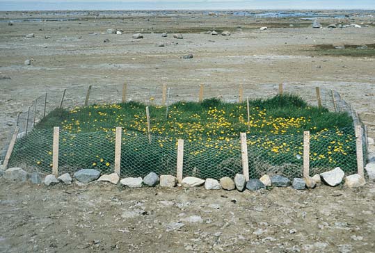
Three decades ago, this positive feedback—birds grazing and defecating on the marsh, the marsh regrowing rapidly, the birds eating and adding nitrogen, again—resulted in increased above-ground primary production compared to ungrazed marshes. In other words, the birds strongly influenced plant growth in the salt marshes. One of the authors (Jefferies) and his colleagues constructed enclosures in the intertidal salt marshes to keep out geese, and primary production declined within a season. In the past 20 years, though, the impact of this positive feedback declined dramatically. Something changed.
According to a U.S. Fish and Wildlife Service survey 30 years ago, 600,000 light geese—mostly lesser snow geese—turned up in a mid-winter count. Today, such a count reveals 3 million birds, and the actual population size is probably at least double that. Traditionally, the mid-continent population of lesser snow geese fed in winter on the above- and below-ground tissues of salt-marsh plants in the coastal marshes of Texas and Louisiana. From the 1940s onward, snow geese began to feed on spent rice in the prairies adjacent to the Gulf salt marshes. The big changes came between the 1960s and 1980s, coincident with the introduction of high-yielding crops and increased use of nitrogenous fertilizers. Geese started eating other crops—corn, soybeans and wheat—during the spring and fall migrations. In addition, many birds no longer wintered in the Gulf states, but remained in other southern states, including Arkansas and Missouri, foraging on agricultural crops.
In the past, the availability of food in the Gulf salt marshes limited the size of the mid-continent population of geese. Now, with geese feeding in agricultural fields during the winter, limitations in food do not hold down the population size. Other factors, too, including food supplements put out at refuges, may add to the population growth for geese, but the agricultural bonanza seems to explain most of the increase. Indeed, similar changes in agricultural practices in the Netherlands triggered long-term shifts in the abundances and the feeding habits of ducks, geese and swans.
After a winter of good eating, the geese fly north. In early spring, immediately after the ground has thawed but before the above-ground growth of grasses and sedges begins, the birds dig for the roots and rhizomes of these salt-marsh plants. An adult goose can remove everything—the vegetation above and below ground—across one square meter in just an hour. With three million geese, that translates to stripping three million square meters every hour. Both lesser snow geese and Canada geese contribute to this destruction.
Pete Saloutos / Corbis
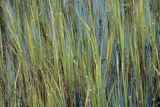
Damage also develops in the freshwater marshes of the Hudson Bay Lowlands. There lesser snow geese pull up the shoots of sedges in spring, eat the base, which is rich in nutrients, and discard the rest. Large rafts of destroyed shoots float on meltwater in these marshes in early spring. Successive episodes of shoot-pulling each season weaken the sedge plants, and they die, exposing the underlying peat surface. So far, waterfowl attacks have destroyed about one third of the Hudson Bay Lowland's coastal salt-marsh vegetation. Another third shows severe damage, unlikely to recover in the presence of geese. Geese also graze heavily on the remaining third.
This grazing causes a second positive feedback loop. At high populations, the geese can destroy a salt-marsh sward, or grassy covering. Removing the sward exposes sediment in an intertidal marsh, which sets off increased evaporation. Removing more water from the soil increases its salinity. In the upper layers of sediment, right where the roots of the grasses and sedges grow, the salinity can reach three times that of seawater, making it inhospitable for recolonization. Without the influence of established vegetation shading the surface, not only does the soil salinity increase, but the soil also becomes anoxic because diffusion of oxygen from plant roots does not occur.
Photograph courtesy of Robert Jefferies.
Salty and anoxic soil prevents plants from recolonizing exposed sediment, at least in the short-term. Then, erosion quickly removes the thin veneer of organic soil, and the peaty substrate starts to oxidize. In combination, that destroys the seeds normally left from the growing grasses and forbs. Worse still, the two most common plants, a grass and a sedge, are either sterile or flower very infrequently. Consequently, only clonal growth—a slow process—from remaining vegetation could fill in bare spots. Unfortunately, the grubbing geese create exposed patches, which spread and eventually come together as bare mudflats. Despite the positive impact of goose excrement, the root digging wins, leaving nearly irreversible changes in soil properties of exposed sediment.
Even if grazing pressure were to subside, only decades could bring a full recovery. In any case, grazing pressure continues, and the degraded areas in the coastal landscape of the Hudson Bay Lowland continue to spread. The severe destruction of the salt marshes removes an important resource for many organisms, including birds, terrestrial and aquatic insects, and soil microorganisms. The damage could spread even more. Other Arctic habitats, including inshore marine ecosystems that are heavily utilized by seals, fish and polar bears, may also be put at risk by the sharp decrease in the production base of the intertidal flats—all because of an agricultural abundance 5,000 kilometers to the south.
The geese of the Hudson Bay Lowland challenge one of the most widely held beliefs about the dynamics of salt-marsh ecosystems: Supposedly, consumers do not play a large role in controlling primary productivity of marsh systems; rather, marshes are controlled exclusively by resources, or bottom-up processes. In both positive-feedback systems described here, however, geese do regulate primary production.
For another example of consumer impact, we turn to the marshes of Georgia and the Carolinas—long known as some of the most productive natural systems on the planet. Cordgrass dominates daily flooded portions of these marshes as dense monocultures, and it accounts for most of the primary production of these ecosystems. Nearly half a century ago, seminal work on this system on Sapelo Island at the University of Georgia Marine Institute by Eugene Odum, John Teal and their colleagues characterized these systems as strongly controlled by physical factors and nutrient supplies, with consumers playing an insignificant role. Odum and his colleagues considered cordgrass unpalatable to most consumers and proposed that it entered the food chain as dead shoots. Most ecologists accepted the detritus-based model of a marsh food web, and even extrapolated it to other marine habitats, including mangrove and seagrass ecosystems. Although this model assumed an insignificant role for consumers, that claim was not rigorously tested until recently.
On Sapelo Island and other marshes on the southeastern coast of North America from Virginia to the Gulf of Mexico, the marsh snail (Littoraria irrorata) is one of the most conspicuous and abundant invertebrates. Ecologists have long considered Littoraria as a detritivore specialist that consumes the microorganisms and fungi that attack dead and dying cordgrass. During low tide, Littoraria browses on organic matter on the marsh surface and, at high tide, ascends cordgrass to feed on standing, dead Spartina and its associated microbes. On Sapelo Island, this snail often exists in extremely high densities—500 to 1,500 individuals per square meter—on the terrestrial border of the cordgrass zone, where marsh grass is severely stunted. It is particularly common to find this snail in discrete "die-back" zones, where the majority of plants are dead or dying.
Early marsh ecologists assumed that harsh physical conditions killed cordgrass in these die-back areas, which in turn attracted large numbers of detritivore snails. Current work by one of us (Silliman) and collaborators, however, revealed that this snail actually grazes live cordgrass and, in the process, exerts strong control on marsh-plant growth. In areas of high snail density, extensive grazing scars—longitudinal grooves made by the snails—exist on cordgrass blades. At low densities, fewer than 50 snails per square meter, Littoraria grazes primarily on fungus that lives on dead marsh grass. At moderate to high densities, 50 to 1,500 individuals per square meter, snails cultivate crops of nutritious fungi directly on the surface of live cordgrass leaves. The grazing scars on live cordgrass get infected by marsh fungi, whose spores are ubiquitous across the marsh surface. Snails further enhance fungal crop growth in grazing scars by depositing nitrogen-rich fecal pellets and actively graze these fungus-infected wounds for nutrition. The combination of fungal infection and snail grazing kills the scarred leaves, and can completely kill cordgrass—all of the material both above and below ground.
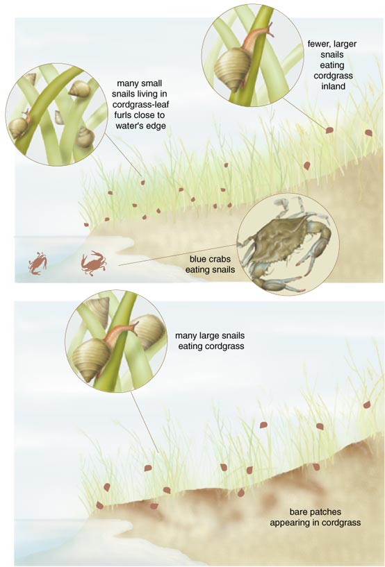
Two of us (Silliman and Bertness), excluded snails from high-marsh areas on Sapelo Island, and that experiment created an order of magnitude increase in cordgrass production, showing that snails naturally suppress this grass. The impact of snails proved even more dramatic in the tall, productive grass on the seaward border of the marsh. When we added moderate densities of snails there—where snails are typically absent—they entirely destroyed the cordgrass in fewer than eight months. In other words, snails converted one of the most productive ecosystems on the planet to a barren mudflat.
Typically, other consumers keep snails away from the seaward border of marshes. Juvenile snails live in the leaf furrows on productive, tall cordgrass on the seaward border of these marshes, but crabs, fish and turtles eat the young snails once they grow too large to fit in the furrows. As a result, few mature snails survive on the productive cordgrass at the marsh edge. At higher marsh elevations, snails increase in number because the dense wall of cordgrass blocks large predators, such as blue crabs.
These results suggest that a so-called trophic cascade produces the highly productive marshes of Georgia and the Carolinas. That is, crabs and other predators eat the snails that eat the fungus that infects the cordgrass, which makes up much of these salt marshes. Without snail predators, increasing snail densities could decimate these marshes. Ecologists long considered trophic cascades important in structuring subtidal kelp-bed communities and temperate-lake plankton communities, but not vascular-plant communities.
Sea otters of the Eastern Pacific kelp communities make up a well-known example of a trophic cascade. The sea otters keep populations of sea urchins in check, preventing them from suppressing kelp production. When fur traders harvested sea otters to near extinction for their pelts at the end of the 19th century, sea urchin populations grew rapidly and decimated the kelp forests, leaving bare substrate covered only with unpalatable calcareous algae.
A similar relationship exists between Littoraria, its predators and southern marshes. Littoraria could decimate these marshes, but predators keep snail populations at low densities, which results in productive marshes. Today, though, this cascade appears out of balance. When commercial fishers take heavy catches of blue crabs—one of the most voracious predators of Littoraria—and crab populations are simultaneously hit with disease and salinity stress from drought, extensive diebacks have appeared in southeastern salt marshes. These findings warn that overharvesting blue crabs may be an important contributing factor to marsh dieoff by releasing Littoraria from consumer control, which would lead to increased snail populations that can destroy cordgrass. In addition, the fact that highly productive cordgrass suffers the most damage from snail grazing warns that nitrogen loading, a pervasive problem in Atlantic-coast marshes, may increase the vulnerability of marshes to snail attacks.
As these examples reveal, salt marshes all along the Atlantic coast face a hazardous future. Marsh ecosystems suffer from shoreline development in New England, changing agricultural practices in the south-central United States and overharvesting of blue crabs off the Carolina shores. These problems, and others, which are occurring worldwide—intertwined as they are with human practices of development and consumption—will be difficult to solve. Working them out, however, is the only way to salvage the many services provided by these ecologically important habitats. To do so, we need a predictive, mechanistic understanding of the factors that maintain the structure and organization of these systems, and this information needs to get into the hands of proactive conservation groups, the general public and responsible politicians. If this doesn't happen quickly, we might lose most of the remaining salt marshes, especially those on the Atlantic coast.
Click "American Scientist" to access home page
American Scientist Comments and Discussion
To discuss our articles or comment on them, please share them and tag American Scientist on social media platforms. Here are links to our profiles on Twitter, Facebook, and LinkedIn.
If we re-share your post, we will moderate comments/discussion following our comments policy.