
This Article From Issue
January-February 2004
Volume 92, Number 1
DOI: 10.1511/2004.45.0
In a Perfect Ocean: The State of Fisheries and Ecosystems in the North Atlantic Ocean. Daniel Pauly and Jay Maclean. xxxii + 175 pp. Island Press, 2003. Paper, $25.
Because more than 60 percent of the Earth's swelling population lives within 100 kilometers of the seacoast, it's not surprising that the litany of human insults to the oceans is extensive and growing: Pollution, habitat destruction and degradation, importation of invasive species, and fisheries have all had devastating effects on marine biodiversity. Daniel Pauly and Jay Maclean's In a Perfect Ocean reports the findings of an ambitious project that undertook to measure the harm fishing has inflicted on the ecosystems of the North Atlantic and to propose ways of reversing the trend. This case study is just the first in a series of investigations that will be carried out by The Sea Around Us Project (www.saup.fisheries.ubc.ca), which the Pew Charitable Trusts sponsors and Pauly, a prolific and influential fisheries scientist, leads. He and his team members have published several high-profile papers since 1995, and these articles are forcing changes in our views of how fishing influences marine ecosystems around the world. For instance, the team's analyses reveal that humanity already demands a preponderance of the ocean's productivity. Pauly's work is controversial and has been the subject of considerable criticism charging that he oversimplifies complex issues and misinterprets fisheries statistics. Despite this, his efforts have been instrumental in changing the focus in management of marine resources from single species to complete ecosystems.
Ad Right
The findings reported in the book will not surprise readers familiar with Pauly's research. However, the consolidation of his earlier inferences into one treatise focused on a single ecosystem makes a compelling, indeed startling, case that the North Atlantic Ocean is severely degraded and remains under attack.
After nicely setting the stage in the introduction, Pauly and Maclean concisely chronicle the North Atlantic and its long history of exploitation. This account is interesting and significant in its own right, but here it furnishes the appropriate baseline against which to gauge the effects of fishing. Although Pauly and Maclean compare the current situation with that in 1950 (the first year for which standardized catch statistics are available), in the first chapter they make it clear that by 1950 North Atlantic fishes had already been severely affected. They thus argue that our expectations of what a healthy ocean should look like ought to be based on the historical and paleoecological record.
The authors used software called Ecopath with Ecosim, along with statistical interpolation, to make their assessments. More specifically, they took food-web models covering one-third of the North Atlantic and combined them with spatial catch data, and maps of physical features, covering the entire North Atlantic. This enabled them to estimate fish biomass in the two-thirds of the North Atlantic for which no biomass data were available.
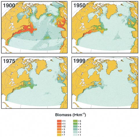
From In a Perfect Ocean.
The book captures the heart of these analyses in two sets of maps that depict dramatic increases in the intensity of fishing and corresponding declines in fish biomass during the 20th century. Next, Pauly and Maclean show that commercial fishers have systematically overharvested the largest and most economically valuable species and thus are now capturing fishes lower on the marine food web than those they were catching 50 years ago. The few remaining large fishes and marine mammals must also feed at a lower level on the web. The authors argue that fishing, by shortening food chains, exposes these top predators to environmentally driven fluctuations in the supply of plankton at the base of the web—fluctuations that were previously damped in more-complex food webs. The dramatic variation in plankton abundance has proved hard to predict, which has made the size of fish stocks hard to predict, which in turn has made fisheries harder to manage than they would be otherwise.
After exposing the roots of the trouble, Pauly and Maclean attempt to describe how the North Atlantic reached its current sorry state. They document several problems—government subsidies that distort the economics of fishing, ineffective governance at both the national and the international level, and a lack of scientific information—all of which, they contend, interact to produce a management regime at odds with ecological reality. They conclude by recommending five measures to restore the North Atlantic: Reduce fishing effort by a factor of three or four; establish 20 percent of the ocean as marine reserves by 2020; increase market-based attempts (such as eco-labeling) to move the fishing industry toward sustainability; implement procedures to expose unsustainable and illegal practices; and alter access and property rights in fisheries to favor small-scale, place-based operations.
Fisheries scientists will find much useful information in this volume. Although it reiterates results Pauly and his colleagues had previously published, the book contains several kernels of ideas that I suspect will stimulate more detailed analyses. For instance, Pauly and Maclean briefly mention "allowable fishing areas," which are the mirror opposite of marine reserves. An approach that focuses on where we should harvest fish offers a different and potentially valuable perspective on the controversy about the efficacy of "no-take" marine reserves as a fisheries management tool. That is, Pauly suggests that it would be better to ask where we could fish without severely degrading marine ecosystems, rather than asking what areas we should avoid in order to ensure the integrity of fish stocks.
Pauly and Maclean's broad-brush approach is suitable for a general audience of the conservation-minded. However, those in the field may be frustrated by the book's sparsity of detail, which makes it difficult to evaluate the authors' conclusions. Readers who want more beef are burdened with turning to the endnotes, which often contain valuable information or literature references.
Ultimately, this treatise is a passionate exposé. Some will surely cavil with its findings or recommendations, but most will agree that the state of the North Atlantic is dismal. How exactly to respond to the problems will be a matter of much debate, but the book makes it abundantly clear that something must be done.—Phillip Levin, National Marine Fisheries Service, Northwest Fisheries Science Center, Seattle
American Scientist Comments and Discussion
To discuss our articles or comment on them, please share them and tag American Scientist on social media platforms. Here are links to our profiles on Twitter, Facebook, and LinkedIn.
If we re-share your post, we will moderate comments/discussion following our comments policy.