Skip to main content
Close
Search
Help
Topics
Features
Blogs
Video
Podcasts
Magazine
Archive
Subscribe
Newsletter
About Us
Advertise
Login
Register
Facebook
YouTube
LinkedIn
Bluesky
American Scientist
Open navigation
Search
Help
Login
Department
Science Observer
Arts Lab
Books
Cartoon
Computing Science
Engineering
Essay
Ethics
From The Editor
From the President
From The Publisher
Hypothesis
Infographic
Letters To The Editor
Macroscope
Marginalia
Perspective
Policy
Science and Engineering Values
Science Communication
Science Observer
Scientific Method
Sightings
Spotlight
Technologue
Finding the Rules that Work
Richard Fiene
Science Observer
Policy
Social Science
Statistics
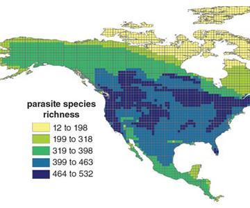
Extinction Spillover
Katie L. Burke
Science Observer
Environment
Ecology
Even Birdbrains Learn from Experience
Jenny Jennings Foerst
Science Observer
Biology
Animal Behavior
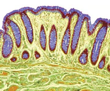
Night Shift Belly
Jenny Jennings Foerst
Science Observer
Medicine
Physiology
In the News
Science Observer
Communications
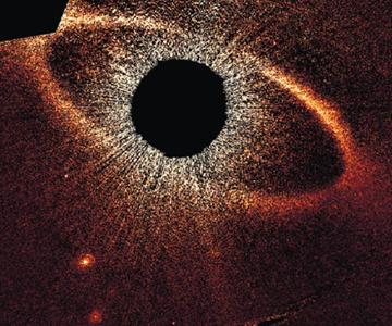
Pulling On the Shade
Fenella Saunders
Science Observer
Chemistry
Physics
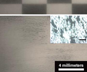
Tiny Lenses See the Big Picture
Fenella Saunders
Science Observer
Physics
Technology
Photography
Load more
×
AMSCI ICON NAVIGATION:
Navigation Menu
Search
Help
Log In, Register
My AmSci
Select Options
(not present on all pages)
Click "American Scientist" to access home page
×