Deconstructing DNA Beyond the Helix
By Caryn Babaian
An artist’s experimental approach to Rosalind Franklin’s Photo 51 reveals the molecule’s intricate biochemistry.
An artist’s experimental approach to Rosalind Franklin’s Photo 51 reveals the molecule’s intricate biochemistry.

In 1952, British chemist Rosalind Franklin bombarded crystallized DNA with x-rays and recorded how the rays diffracted through the sample. The result was Photo 51, one of the most astonishing and iconic images in science. As the x-rays bounced off different parts of the DNA, they created patterns that captured the shapes within the molecule. To the trained eye, Franklin’s image showed that DNA has a double helix shape, a realization that transformed our understanding of modern biology.
The repercussions of this discovery may be best appreciated by molecular biologists, mathematicians, and physicists, but artists who study the science can unveil new ideas, too. While scientists label parts and define quantities in experiments, the artist’s mind searches for patterns, experiencing them as organic, neuronal, visceral, and sensory imprints.
Today, DNA is universally identifiable as a highly stylized and graphically modified double helix icon. As soon as we see a twisted ladder, we know that it is meant to represent DNA. There is even an emoji for DNA. To researchers and the layperson alike, the helix of DNA is generally regarded as a one-perspective phenomenon: It is the genetic code, biological heredity expressed in a single geometric form. Nevertheless, there are many other ways to look at DNA, and perhaps many other shapes to be noticed once we shift to different perspectives. As we will see, the molecule’s interaction with water can change the twistiness and structural characteristics of the iconic helix.
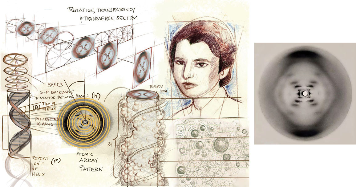
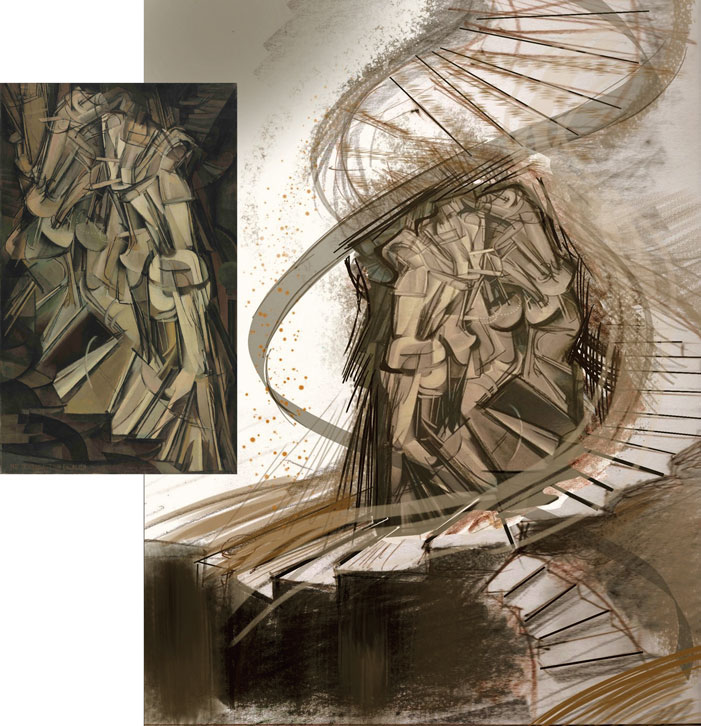
An illustration by the author (above) depicts Rosalind Franklin’s process in 1952 as she caught DNA on film for the first time using x-ray diffraction, in the now iconic Photo 51 (right). This X-shaped image shows the molecule in cross-section. Each black mark in the X represents a different level in a stacked array of planes. The two mirror image sides of the X represent a complementary set of stacked nucleotides, and the distance between each black mark indicates the distance between each helical turn. The hash marks toward the margins have more space in between them than those near the center, which showed that the molecule was made of two strands.
All images courtesy of the author, unless otherwise noted; image on right from Franklin & Gosling, Nature, 1953
One point of view is Franklin’s Photo 51, the forbearer of this molecule’s fame. Franklin organized DNA strands around fibers and then used x-rays to slice through the then-mysterious molecule, capturing a perspective of it in cross section on film. Photo 51 enabled James Watson and Francis Crick to elucidate the double helix into a tangible model. But it was still just that: a model. A key to capturing an image of DNA was holding it still. But DNA is never still in real life—it’s fluid and dynamic.
When one considers the leap of imagination from the X shape in Photo 51 to the helical model, one gets a feel for the many layers of meaning within the original image. As both a scientist and an artist, I began revisiting Franklin’s experiment through drawing (above image), to hone a visceral feeling of her process and to raise both new and old questions about the shapes of DNA, as well as its interactions with the aqueous space inside the cell’s nucleus, which often transforms DNA’s functionality.
DNA is not an isolated molecule. It is shifting and changing all the time. It is sheathed by layers of water. How does it look in this watery sheath? What do the transitional shapes of DNA look like when it forms affiliations through a complex vibrational lattice of hydrogen bonds with enzymes and ligands? What does DNA look like when its watery coating swells or shrinks?
For me, understanding these processes has rekindled a sense of wonder for the “blueprint of life,” the molecular instructions for making a human or any other living thing. I now appreciate more fully nature’s hidden and astonishing capacity for generating endlessly beautiful forms. DNA, commonly depicted as a simple twisted ladder, in fact has a multifaceted life of its own. An artistic approach is not only aesthetic and emotional, but also a way to understand DNA as a dynamic, complicated, and still-mysterious molecule at the heart of the living world.
Process is experience, an embodied familiarity with action, change, and relationships. Stable molecules, phenotypes, and ecosystems are in flux, not finished goals. The discovery of DNA was the culmination of multifaceted processes performed by many individuals. Process is a universal action in every living thing—and in the molecules that keep us functioning—and process is also a human action, such as designing and executing an experiment. Focusing not only on static images of a molecule, but also on these processes, leads to a far deeper understanding of life and the molecules that sustain it. Although it is difficult for most people to interpret Photo 51, Franklin’s notebook reveals her planning and conceptualizations about the x-ray diffraction process.
Franklin’s experiment began at King’s College London in 1951. She used a salt solution to maintain a small aqueous component around fibers of DNA. She then positioned the fibers in a parallel arrangement and suspended them from a paper clip in the pathway of emitted x-rays under controlled humidity. The x-rays were accelerated into the DNA lattices and interacted with the geometry of the planes of the DNA strands. When the x-rays passed through the sample, they diffracted at angles off the bases at the center, which were exposed on a piece of photographic film, resulting in a relatively uniform pattern of lines that created the now-famous X shape.
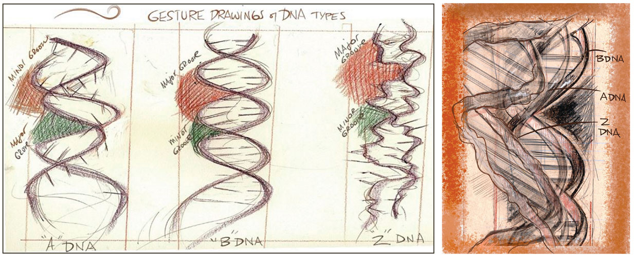
Franklin’s work helped define two different forms of DNA, the A and B forms, both of which are right-handed double helices. The B form is the most common, and the A form is more compact, with a wider major groove and a shorter helical structure. In the B form of DNA, the “rungs” of the twisted ladder are straight across—perpendicular to the helix axis. In the A form, the rungs are angled across the helix axis. In Franklin’s images, humidity affected the form of the molecule. The A form was seen at 0–75 percent humidity, and the B form was seen at 75–100 percent humidity, with the differences between the two helices measured in angstroms—smaller than a nanometer.
The X shape that Franklin observed was not made of solid forms, but of fuzzy dark lines, fuzzier toward the margins and darker and more defined toward the center. If one works back from the helix we know today to the cross-sectional X-shaped image in Photo 51, one can approach the helix as an array of planes at repeating angles, stacked upon one another. Each black line represents a repetition of atoms. The two mirror image sides of the X represent a complementary set of stacked nucleotides. (See the figure above, on the right.) The distance between each black mark indicates the distance between each helical turn—for the B form, 34 angstroms. The distance between the center and the dark shapes at the top and bottom of the image can be used to calculate the distance between each ladder rung—3.4 angstroms in Photo 51’s case. The hash marks toward the margins have more space in between them than those toward the center, which indicated that the molecule was made of two strands.
In the 1950s and 1960s, x-ray diffraction images were difficult to create, taking several days at a time and exposing the experimentalist to radiation. But in this lag before the image was developed, Franklin enjoyed an incubation period of sorts, during which she could marinate ideas carefully in her notebooks. Would the same level of understanding and artistic eye for the helix shape have emerged without that slowed time? This “slow” gestational time is also process, and it is a shared characteristic of both high-quality, thoughtful science and thorough, investigational, attentive art. Franklin participated in activities outside of her work too, such as hiking and attending concerts; being in nature or experiencing music or art can help refine perspective on a problem.
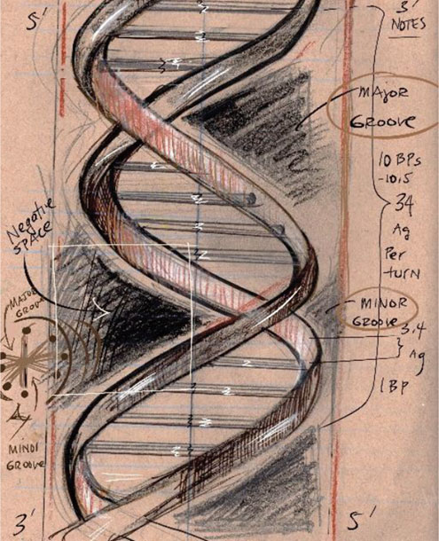
Rotation, transparency, and transverse sections—illustration techniques popularized by Leonardo da Vinci—have long been used in science to portray the complex geometric clues held in every black-and-white, two-dimensional image. These illustration methods help us envision matter on multiple planes and solve visual problems. It is not just structure or geometry that is necessary for these interpretations, but also an appreciation of gray scales and of the subtle changes and variations in x-ray film.
In the long tradition of artists doing portraiture or landscapes, gray scales often map out a range of tonal values. Creating tonal values aids in understanding transparencies and three-dimensional shapes. These tonal values may correspond to the amount of light, the direction of a light source, or the curvature of an object. They can also help depict three-dimensionality. Even though DNA exists at the molecular level and is usually not exposed to any light, these same techniques can help depict its structure and dynamics. Typically done with charcoal, pencil, or oil paint, creating gray scales with different media trains the eye to recognize and feel value differences.
Popular depictions of DNA typically present it as a static object. Each repeating unit of DNA has three linked entities: the phosphate group, the deoxyribose sugar, and the bases. In the double helix’s twisted ladder, the rungs of the ladder are the base pairs—the nucleotides adenine, cytosine, guanine, and thymine represented by the letters A, C, G, and T—forming the genetic sequences that code for proteins and include other molecular instructions. The spiraling sides of the ladder are known as the sugar-phosphate backbone, the structural support for the molecule.
The classic image of DNA focuses on what the nucleotide sequences code, which makes DNA’s functionality appear one-dimensional. But considering the positioning and movement of DNA’s constituents adds another perspective. With drawings, one can animate DNA dynamics and atomic-molecular interfaces, creating models of deep atomic caves, where much of the real action happens. Drawings can explore those possible changes, because employing da Vinci’s illustration techniques can help stabilize the view. One might envision staggered rises, shifts, and slides occurring within DNA as it unzips or interacts with proteins, or as it puckers and vibrates through its sugar-phosphate backbone.
Two grooves, one wider than the other, form on either side of the helix, known as the major and minor grooves, respectively. These gaps between the rungs’ twisted strands are formed by the bonding of the sugar molecule in the backbone, deoxyribose. Using da Vinci’s rules, one can picture a nucleotide, such as adenine (A) or guanine (G), tilted along the x-axis, clarifying the major or minor grooves and drawn with angular characteristics. A propeller twist maintains the staggered nucleotides in a more ridged position and displaces them slightly. Maybe these rolls, twists, and tilts are sequence dependent, leading to a better understanding of function.
One can also use negative space to appreciate that, as the name implies, the major groove is wider than the minor groove and that many proteins bind in the major groove, a fluid but stable depression with stronger binding affinities. The differences in the grooves of each helical turn are more noticeable when one applies negative space. By comparing negative and positive space, we notice more topological features.
DNA can also take many forms (above). Molecular biologists estimate that there are at least seven forms of DNA, maybe more. A and B forms were discovered first, but D and Z forms have been studied as well. All forms except the Z form are said to be twisted in a clockwise direction. In DNA’s varying configurations, conformational details can determine how mutations are introduced into a sequence and whether or how they are repaired. These configurations can even determine whether the sequence is functionally altered by its current shape. As form changes, so does function.
Drawing or illustrating a DNA molecule allows one to examine the molecule’s bonding, relationships, and structure closely, and to contemplate its nature.
DNA’s 3D configuration merges with the interior of the cell. DNA is constantly moving, and as bases pair and protons migrate, its 3D structure is dynamic. The speed of these changes is so fast and tiny that they are unlikely to be witnessed in DNA’s nuclear home, nor captured in an x-ray diffraction image. DNA replication occurs at speeds 100 times faster than previously predicted—thousands of base pairs per minute—making the nucleus a busy but organized studio of cellular strategies.
The remarkable stability of the DNA molecule is necessary as protons are transferred through quantum tunnels in its watery world, with an occasional hydrogen proton forming a rare tautomer. The movements are so small and fast that there are moments when DNA may not even be a material object. To depict this speedy epicenter, we need an artist’s perspective. Do some configurations and transitional states trigger a higher propensity for mutation? Such questions are at the forefront of biochemistry research, and illustration is a necessary part of that endeavor.
DNA is normally depicted as an isolated molecule, but that is never true. The entire surface of a DNA double helix is coated in layers of water molecules. This sheath of water attaches to the genetic material through hydrogen bonds, made by sharing hydrogen atoms between molecules. Through these hydrogen bonds, water can influence how DNA takes shape and interacts with other molecules. Change is constant, and DNA is in a comingled relationship with the lattice of water. In some cases, water assists proteins in recognizing DNA sequences.
“Water serves as a mediator between DNA and other molecules, even for very specific interactions,” says biochemist Martin Egli of Vanderbilt University. “Before any molecule can bind to a segment of DNA, it must first go through this water shell.”
While in its watery niche, a base stacks and twists along a plane as molecular forces repel or attract it ever so subtly. Electrons in uncharged molecules can cause weak interactions with nearby molecules called van der Waals forces, which are often but not always transient. (See Little Interactions Mean a Lot, March–April 2014.) Positively and negatively charged ions pull toward one another through coulombic attraction. These pushes and pulls challenge our conception of the standard DNA form. Bases move, the backbone moves, and there is a kinetic flow, a charged gesticulation that reverberates. The negative charge of the backbone is a platform for sequence-independent electrostatic interactions with proteins in the cell. The DNA molecule is but a temporary relationship of energetic musicians and ballerinas carrying out their roles, adjusting and ad-libbing accordingly on a cellular stage filled with water.
DNA balances its intramolecular and intermolecular interactions to maintain homeostasis. Biology education typically focuses on the sequences of bases, whereas the sugar phosphate backbone goes largely unnoticed. Biology classes emphasize the linear process of copying selected segments of DNA, transcription and translation, and the expression of genes that ultimately leads to the production of proteins. Yet DNA is changing, all over. One interaction can prompt change throughout the molecule. Could the backbone be influential in gene expression? It might depend on how that action reverberates through the molecule.
Water creates a cozy environment for DNA, so that bonds form in strong, stable ways. Water promotes base stacking, when base pairs snuggle up together through hydrogen bonding. Water also solvates the surfaces of the major groove, forming well-defined hydrogen-bonded networks that bridge the two strands across the minor groove. The infrastructure of life is wet. When biology students are memorizing the parts of DNA, I encourage them to consider how those parts got there and what each part interfaces with. Down to the last molecular pirouette, the angles around the bonds that hold two nucleotides together are engaged in a world of alterations that can affect the way the entire DNA molecule functions in its moist arena of cellular activities.
Life originated in water, and each living organism still carries a watery chemical environment within. When a discipline is defined by a molecule or makes it a central dogma, we might use artistic thinking to explore the protean attributes, its close associations, a little further. There doesn’t appear to be math for all this interconnected transiency in DNA; there is no overall vacillation potential for DNA, no interconvertibility quotient, no water mingling coefficient. Art can refine these ideas. Biologists studying this topic work with dynamic, highly specific, short-lived, moment-to-moment relationships. Their models are attempts to understand this complexity, which can be informed by artistic visions coupled with biologists’ experimentation. For the abstract concept of DNA, art can help realign our thoughts about living systems as we scale up its flexible and replicative potential to the cellular level, and then to the phenotype.
Franklin’s experiment also highlighted the importance of water regarding the B form of DNA, to which water molecules cling, making the DNA molecule flare out. Through illustration, we can start to imagine how bases in that rigid helix might change as a result of the backbone’s dynamics, how the DNA might convert from B to A or Z forms, and whether important unknown biological functions exist in intermediate states in various associations with water.
For this out-of-focus, vibrational perspective—that is, these intermediate transitions—we might look to the artist Marcel Duchamp and his infamous painting Nude Descending a Staircase.
A painting can be static but also dynamic in meaning; it can provide a familiar way to interpret the dynamic nature of DNA, even though it is technically stationary imagery. The quantitative scientist calculates technical details—pi bonding, dynamic equilibrium coefficients, distance measurements per base, and putative stacking interactions. If scientists want a better image of this structure, they can refer to a file on torsion angles or a computational program on atomic simulations. Or they can look at Marcel Duchamp’s Nude Descending a Staircase. (See the images below.)
Marcel Duchamp’s 1912 painting Nude Descending a Staircase (left) depicts motion, rotation, and transparency, as the nude slips from one transitional form to another. This painting technique could also suggest the wobbly, flexible quality of DNA (right), along with the overall stability of its twisted ladder structure.
Philadelphia Museum of Art/Wikimedia Commons
Duchamp’s famous painting is a depiction of motion, rotation, transparency, and the same form (the nude) slipping into one transitional form after another. The painting presents both the original nude and the nude after a few moments, captured in her dynamic state. Vibrations in each step transmit to the next, down the staircase. One might say the painting is demonstrating van der Waals forces between the nude and the stairs. Perhaps this connection is not surprising, as the picture was created at the precipice of the discovery of quantum mechanics. Looking at Nude Descending a Staircase, we see superposition of form. In this way, the intermediate figures create an out-of-focus image as adhesion and entanglement exist between the figure and her associated staircase, and within the figure herself. In the many fleeting images, there is both only one figure and many figures at the same moment.
Van der Waals forces are like an invisible rope holding a wooden bridge together; mutable, electrostatic polarizations between the vibrating subdivisions of the bridge make one and many moments the same and different, yet still held together in an identifiable form. One could try to develop empirical methods to study the properties of these forces and how they influence DNA structure, or one could develop a theoretical model of the perceived energy landscape, calculating relative stabilities within this wobbling bridge. When a misstep occurs in the descent down the staircase, Duchamp’s nude might experience a slight twist of an ankle resulting in shifts in the torsion preferences from one step to another, and this misstep might correspond to changing orientations of the bond that links the bases to the sugar-phosphate backbone. In that tiny gaffe, the mutagenicity potential might be indirectly affected. This visual story is similar to DNA’s story: a misstep, an awkward entanglement, a mutation, a repair, a hydrated spine that follows the flow of its watery world.
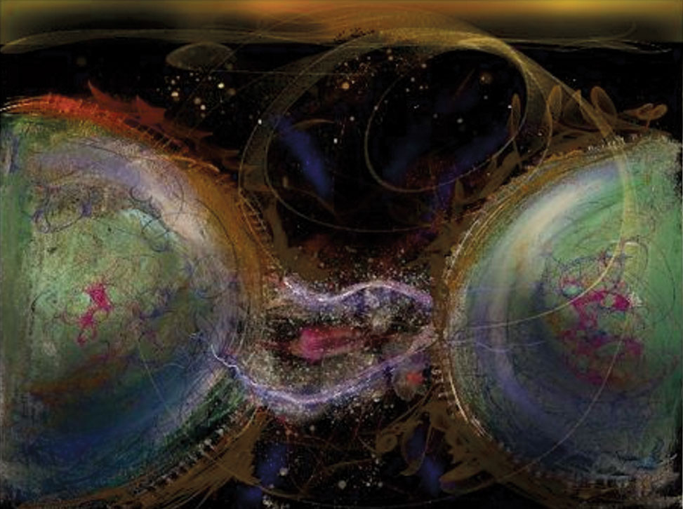
This conceptual image of van der Waals forces shows how electrons in uncharged molecules can cause weak interactions with nearby molecules. These forces are like an invisible rope holding a wooden bridge together.
Duchamp’s painting helps us see how in the blurred motion, there is a form and forms—conformational change. The painting fosters an appreciation for structural dynamics, without the need for the calculations of quantitative study. The nude is an electrostatic model; it is simulating variations of movement from point A to point B. The nude depicts a wobbly, flexible quality to DNA among the molecule’s overall stability. Like the vibrating DNA and its watery shell, the nude and the stairway are sometimes one.
Life originated in water, and each living organism still carries a watery chemical environment within.
Stability is subjective. Stability can be strong, or soft, or in between—just like the bridge, just like a plasma membrane that must remain fluid, but not too fluid and not too rigid. We know that rigid cells are a structural hallmark of diseases such as cancer. From this picture, one may come to see that stability and fluidity are contrasting qualities of conserved biological molecules. These qualities may seem paradoxical, but they are adaptive assets, making life pliable and soft, yet sturdy and stable through time.
Drawing or illustrating a DNA molecule allows one to examine the molecule’s bonding, relationships, and structure closely and to contemplate its nature. While Duchamp’s painting explores the changes of an entire entity, the same effects at the smaller scale of atoms, electrons, and energy might require a different approach. The painting style best suited to this challenge might be pointillism, which uses dots to create brilliant mosaic-like illustrations. Georges Seurat’s A Sunday Afternoon on the Island of La Grande Jatte is a famous example of pointillism. In classical scientific illustration, a similar technique is known as stippling.

Both stippling and pointillism create the feeling of kinetic motion, which underlies the behavior of molecules. Linus Pauling put this technique to excellent use in his work in the late 1940s to visualize how antigens and antibodies bond to one another. Pauling’s artist and collaborator, Roger Hayward, produced many excellent examples of dotted energy relationships. Hayward’s pastel of a molecule (see the image above) does a superior job of conveying the soft, kinetic properties of molecules, while revealing their flexible but resilient lattices. Compare this painting with today’s computer-generated images, which are often hard-edged and lack a sense of movement. Both Hayward and Seurat used their unique styles to capture the dynamism of life, and such approaches could be used to diversify the visualizations of DNA beyond the static textbook helix.
Classical drawing, sketching, and painting are important tools for studying process and pattern. They are time machines, in a way, that can lead one into places that seem opaque and murky. Students of science need to know and experience the practice of skillful observations and the drawings that accompany it. The textures, techniques, and tools of art can broaden our ability to imagine and see into unknown worlds. For DNA, too, there remains a fascinating world of undiscovered intermediates and transient forms and functions, as will always be the case when humans study nature. But with the help of art, we may be able to tunnel into the hidden worlds a little deeper.
DNA is so complex that it can be a challenge to grasp how the parts we learn about connect to the context of DNA in a watery cell of a living organism. The entire molecule of DNA is interactive and has the potential to change as molecules vibrate and bonds bend. Other molecules recognize B-DNA and cooperate with it based on its sequence-dependent properties. DNA can also form shapes and structures that are deviations from its accepted forms, such as guanine quadraplexes, topologically highly variable DNA forms organized through cation-stabilized guanine tetrads, which have pivotal genomic functions.
Visualizing the complex and inconstant nature of DNA demands innovative art that attends to experimental processes and considers the movement and interactions of DNA. Such new visualizations using simple drawings and notebooks could even lead to new hypotheses in molecular biology, because a deep understanding of how DNA interacts in its biochemical, watery environment is at the forefront of many questions in the field of biology and genomics. Franklin dedicated significant time to her notebooks, interpreting her experimental work on paper; perhaps following her example will lead to the next leap forward in the life sciences.
Click "American Scientist" to access home page

American Scientist Comments and Discussion
To discuss our articles or comment on them, please share them and tag American Scientist on social media platforms. Here are links to our profiles on Twitter, Facebook, and LinkedIn.
If we re-share your post, we will moderate comments/discussion following our comments policy.