Skip to main content
Close
Search
Help
Topics
Features
Blogs
Video
Podcasts
Magazine
Archive
Subscribe
Newsletter
About Us
Advertise
Login
Register
Facebook
YouTube
LinkedIn
Bluesky
American Scientist
Open navigation
Search
Help
Login
Tags
Morphology
Aeronautics
Aesthetics
America250
Anatomy
Animal Behavior
Anthropogeography
Archaeology
Astrogeology
Astronautics
Astrophysics
Botany
Cartography
Chronology
Climatology
Cosmology
Crystallography
Cybernetics
Cytology
Ecology
Embryology
Epidemiology
Excerpt
Forestry
Geneaology
Genetics
Geographical Distribution
Geography
Geology
History Of Civilization
Human Ecology
Immunology
Information resources (general)
Information Theory
Linguistics
Logic
Metallurgy
Methodology
Microbiology
Mineralogy
Morphology
Motor Vehicles
Natural History
Nature Conservation
Neuroscience
Oceanography
Paleontology
Petrology
Philosophy of Science
Photography
Physiology
Poetry
Rationalism
Religion
Reproduction
Review
Scientists' Nightstand
Social Science
Special Collections
Statistics
Theology
Transportation
Virology
Zoology
Tardigrades’ Protective Protein
Tyler J. Woodward
Biology
Evolution
Cytology
Genetics
Morphology
Zoology
Spotlight
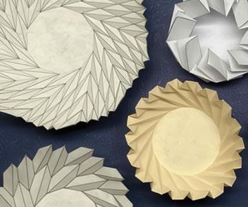
When Art and Engineering Fold Together
Robin Lynn Arnette
Art
Computer
Engineering
Mathematics
Physics
Technology
Aeronautics
Morphology
Sightings
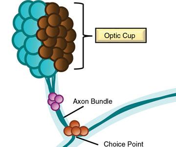
Guideposts for Regeneration
Cyrille Teforlack
Biology
Morphology
Zoology
Spotlight
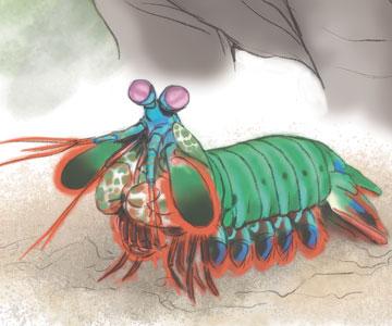
Bioinspired Materials
Rohit Pratyush Behera
,
Hortense Le Ferrand
,
Slocha Sapasakulvanit
Biology
Chemistry
Engineering
Physics
Technology
Anatomy
Crystallography
Metallurgy
Mineralogy
Morphology
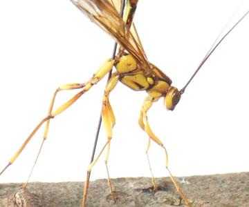
Buckling Without Breaking
Meagan Mulcair
Biology
Engineering
Medicine
Physics
Technology
Anatomy
Animal Behavior
Morphology
Physiology
Zoology
From The Staff
Beautiful Armor
Andreia Salvador
Art
Biology
Morphology
Natural History
Zoology
Load more
×
AMSCI ICON NAVIGATION:
Navigation Menu
Search
Help
Log In, Register
My AmSci
Select Options
(not present on all pages)
Click "American Scientist" to access home page
×